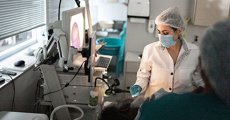
What is Sonography Ultrasound: Overview, Benefits, and Expected Results
The new product is a great addition to our lineup.
Our latest product is an exciting addition to our already impressive lineup! With its innovative features and cutting-edge design, it's sure to be a hit with customers. Don't miss out on this amazing opportunity to upgrade your life!
Sonography, also known as ultrasound imaging, is a medical imaging technology used to create pictures of organs, tissues, and other structures within the human body. This technology is commonly used to investigate the cause of medical problems such as pain or infection, diagnose conditions, and monitor the development of a fetus in early pregnancy. Unlike X-rays, sonography does not use harmful radiation, making it a safer form of imaging.
Unlike X-ray and CT scans, sonography is non-invasive and does not require a laboratory or the aid of a specialist. It uses sound waves to create a detailed image of organs and tissues without penetrating the body’s skin. This is why sonography is considered to be a much safer form of imaging compared to other diagnostic imaging techniques.
## Overview of Sonography Ultrasound
The origins of sonography can be traced back to World War II when it was used to detect submarines using sound waves that could pass through water. In the early 1950s, medical researchers adapted this technology in order to create a more accurate form of medical imaging.
The basic principle behind sonography is quite simple. As sound waves pass through human tissue, like a pregnant woman’s uterus, certain frequencies of sound are absorbed while others are reflected. The sound waves reverberate off the surface of organs and other structures, creating an image for the sonographer to interpret. The image generated by the sound waves is called a sonogram, which is a 2-dimensional picture that can show the size, shape, and position of different structures within the body.
## Benefits and Expected Results
Using sonography, physicians can gain a better understanding of the structure, shape, and size of organs, as well as detect any abnormalities or diseases. Sonography is typically used to diagnose conditions that are not readily visible with just physical examinations or X-rays. The modality is also used to evaluate pregnancies or to assess the condition of a fetus during the early stages of development.
Moreover, sonography is relatively inexpensive compared to other forms of medical imaging, requires no special preparation, and can be done quickly. In addition, sonography is a safer imaging modality compared to X-rays, which use radiation. This makes sonography an ideal choice for pregnant women or other patients who may be sensitive to radiation.
Expected results from a sonography test vary depending on the type of test being done and the condition being investigated. In general, most diagnoses made through sonography can offer immediate feedback, with results made available either during the imaging session or shortly afterwards.
## Case Studies
Sonography has been used to diagnose and monitor many different conditions. Below are two examples of how sonography has been used to diagnose and manage different conditions.
### Follicular Cyst
A 27-year-old woman was referred to a gynaecologist for evaluation of a 5cm pre-ovulatory follicular cyst on the left ovary. The patient underwent a sonography study, which revealed a large, complex-appearing cystic lesion containing macrovesicular cysts, suspicious areas of necrosis, and some edematous masses in the left adnexa. The patient was referred for a laparoscopic cystectomy to remove the cyst and was discharged the same day.
### Uterine Fibroids
A 30-year-old patient was referred to a gynaecologist to evaluate for uterine fibroids. A sonography study revealed multiple small to medium-sized fibroids scattered throughout the uterus, ranging in size from 6-7 cm in size. The patient underwent a laparoscopic myomectomy to remove the fibroids and was discharged the same day.
## Practical Tips and First Hand Experience
When undergoing a sonography procedure, it is important to be comfortable and follow the instructions given by the sonographer. If performed correctly, sonography can provide a comprehensive visualization of the anatomy. Here are some general tips for patients undergoing sonography:
– Dress appropriately for the exam, avoiding clothing with metal or jewelry.
– Ask plenty of questions and voice any concerns you may have.
– Discuss any medications or supplements that you are taking with the sonographer.
– Relax and stay calm during the exam.
From personal experience, I can attest to the accuracy and convenience of sonography. During my last pregnancy, I underwent multiple sonography exams to monitor the development of my baby and to help detect any potential anomalies. Each of the scans provided me with detailed information about my baby’s growth and development, and I was able to visualize the anatomy clearly. The ultrasound provided reassurance that the pregnancy was progressing as expected, with no visible risks or issues.
## Summary
Sonography ultrasound (or sonography imaging) is a safe, non-invasive imaging modality used for diagnosing and monitoring medical conditions. The technology uses sound waves to create detailed images of organs and structures, without the need for laboratory or specialist intervention. Sonography is used to diagnose and monitor conditions such as pregnancy and uterine fibroids, as well as to detect any abnormalities or diseases. The modality is also relatively inexpensive and can provide immediate feedback.
Definition and Overview
Sonography ultrasound, or ultrasonography, is a diagnostic imaging procedure that uses ultrasound waves to visualize different structures inside the body. Ultrasound, a sound pressure wave that oscillates with a higher frequency than what people can normally hear, can measure distance and detect objects. In sonography, ultrasound waves are used to look into the patient’s body and evaluate parts such as blood vessels, joints, tendons, internal organs, and muscles.
Ultrasound is popular as an imaging and examination tool for pregnant women. This procedure is known as obstetric sonography, where ultrasound waves are used to produce real-time images of the foetus inside the patient’s womb. In many countries, obstetric ultrasound is a part of standard prenatal care.
Sonography ultrasound is also quite useful in diagnosing and intervening with a wide variety of dysfunctions, irregularities, and issues in the body. Sonograms can be two-dimensional or three-dimensional, and can display the flow of blood in the veins and arteries, the movement of tissues over a certain period, the location of clots, the presence or absence of certain substances in the body, stiffness or other problems with the soft tissue, and the structure of organs.
Research shows that ultrasonography has a couple of advantages over other kinds of medical imaging procedures. For one, ultrasound images are displayed in real-time, compared to other medical imaging techniques that just take images at a certain point in time, with the images analyzed after they are processed. Ultrasound machines are also portable and can be brought directly to the patient; this is especially useful when the patient cannot be physically moved to a location where an ultrasound machine is located. More importantly, it does not contain radiation, which makes it very safe for pregnant women or very weak patients.
Who Should Undergo and Expected Results
Most laypeople associate ultrasound imaging with pregnancy, as it is a part of standard prenatal care in many countries all over the world. Sonography can indeed be very useful in providing information about the mother’s health and the progress of her pregnancy, as well as improving the accuracy of timing labour because the doctor can see the images of the foetus as it grows. It also reduces the risk of induced labour.
Ultrasound procedures also provide a paediatrician or such specialists with a real-time image of the infant’s brain. Infants are quite delicate and cannot withstand other forms of medical imaging, nor could they follow instructions required by other imaging procedures. Sonography ultrasound can be performed to investigate and diagnose conditions in the eyes, thyroid, bladder, gallbladder, spleen, kidneys, pancreas, testicles, liver, blood vessels, and uterus.
Ultrasound procedures can also be performed while a surgeon is performing a biopsy to monitor and guide the precision and safety of movements.
How Does the Procedure Work?
Before an ultrasound procedure, the patient will be asked to undress and change into a hospital gown. The patient will then be asked to lie down on an examination table for the procedure.
A sonographer, a specially trained ultrasound technician, will apply a lubricating gel on the target area. This gel will reduce the amount of friction between the patient’s skin and the ultrasound transducer, which is a microphone-like device that delivers the ultrasound waves into the skin. The gel is also specially formulated to better transmit and amplify the waves.
The ultrasound waves will echo as soon as they encounter a bone or an organ inside the body, and the echoes will be transmitted back into the attached computer unit.
The patient may be asked to change positions from time to time to analyze different parts of the body. An ultrasound procedure typically takes around half an hour, with the patient perfectly able to return to normal activities after the procedure.
Possible Complications and Risks
Ultrasound sonography is generally safe, without noticeable or serious side effects. This is the reason why pregnant women can safely undergo the procedure. In a study published in 1998, the World Health Organization reported that ultrasound is harmless, and is safe, effective, and very flexible in the provision of “clinically relevant information about most parts of the body in a rapid and cost-effective fashion.”
References:
- American Society of Radiologic Technologists: “Ultrasound.”
- FDA Consumer Health Information: “Taking a Close Look at Ultrasound.” RadiologyInfo.org: “General Ultrasound Imaging.”
/trp_language]
[trp_language language=”ar”][wp_show_posts id=””][/trp_language]
[trp_language language=”fr_FR”][wp_show_posts id=””][/trp_language]


This article is very informative
Nice article–it’s filled with useful information!
This article is very informative
Great post–this definitely cleared up some of my questions about sonography!