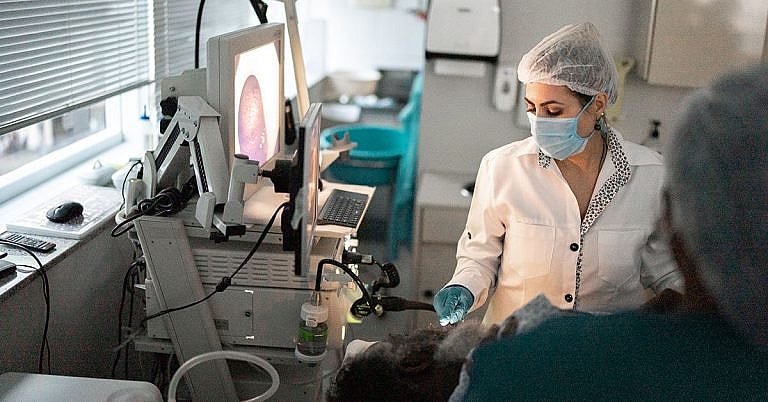
Arterial Doppler
Definition and Overview
An arterial doppler, also known as a vascular doppler or a vascular ultrasound, is a painless and non-invasive diagnostic procedure performed to visualize and evaluate the function of arteries and blood vessels. It creates real-time images of the interior parts of the patient’s body—including the veins, vessels, and arteries that make up the vascular system—through the use of sound waves.
An arterial doppler procedure can effectively and accurately measure blood flow in the blood vessels and the arteries, especially those found in the extremities. Thus, it can be used to detect abnormalities in blood flow and detect the presence of blood clot and poor blood circulation, helping the doctor diagnose related diseases and other medical conditions.
This procedure is commonly used as an alternative to other more invasive imaging procedures, such as venography and arteriography, which both involve the use of dye that is injected into the blood vessels. The dye, also known as a contrast agent, will show up in x-ray images to visualize the network of blood vessels and arteries inside the body. Some people might have allergic reactions to these dyes, as well as other complications. Arterial doppler procedures, which do not use dye, do not have this risk of allergic reactions and complications.
Aside from identifying blockages, clots, and assessing the performance of the vascular system, an arterial doppler procedure can also be performed to:
- Check the condition of the arteries, especially in cases of injuries
- Determine whether a patient is qualified for certain procedures such as angioplasty
- Monitor the progress of treatment plans
- Observe aneurysms and varicose veins to determine the best methods to treat them
Who should undergo and expected results
An arterial doppler procedure is prescribed by doctors:
For patients who are complaining of symptoms such as weakness in the limbs
If the patient is suspected of suffering from deep vein thrombosis
For patients complaining of leg pain
To evaluate the patient’s blood following the development of certain conditions such as stroke that may be causing various symptoms in the vascular system.
To diagnose arterial plaque and determine the severity of the condition
To aid in treatment. Laser and radiofrequency ablation of problematic veins can be guided by a non-invasive imaging procedure like an arterial doppler
To assess and evaluate foetal health. Checking the blood flow passing through the umbilical cord and the placenta can safely check the condition of the foetus’ brain and heart to see if the unborn child is receiving adequate oxygen and nutrients
To confirm if the patient is suffering from superficial thrombophlebitis
Normal results of an arterial doppler procedure mean that the patient’s blood vessels are healthy, and do not have signs of closure, clots, or narrowing of the passages of the blood. On the other hand, abnormal results mean that the patient can be suffering from a condition that needs to be treated at the soonest possible time. These include a blood clot in the arteries, fatty deposits hindering the blood from flowing properly, or dangerous air bubbles occurring inside the blood vessels.
How the procedure works
One of the advantages of arterial doppler procedures is that they do not require a lot of prior preparation. The patient will only be advised to stop the use or intake of products with nicotine, such as cigarettes and chewing tobacco for a half hour to two hours before the procedure. This is because nicotine is known for narrowing the blood vessels, and can thus affect the result of the procedure.
The procedure can be performed by a sonographer or a radiologist, and should be done inside an ultrasound room in a clinic or a hospital. The patient will be advised to remove jewellery or any piece of clothing that can hinder the machine from taking high-quality pictures.
A cold gel will be applied on the transducer or on the patient’s skin to allow the transducer to employ sound waves more efficiently. The sound wave will then strike the arteries, and bounce back to produce real-time images of the arteries on the monitor.
Possible risks and complications
There are no risks and complications involved with an arterial doppler procedure. It is very safe and non-invasive, and can be performed in a short amount of time without the need for hospitalization or long recovery periods. In fact, patients are given a go-signal to go back to their normal activities right after the procedure.
Reference:
- Fowler GC, Reddy B. Noninvasive Venous and Arterial Studies of the Lower Extremities. In: Pfenninger JL, Fowler GC, eds. Pfenninger & Fowler’s Procedures for Primary Care. 3rd ed. Philadelphia, PA: Elsevier Mosby; 2011:chap 88.
/trp_language]
[trp_language language=”ar”][wp_show_posts id=””][/trp_language]
[trp_language language=”fr_FR”][wp_show_posts id=””][/trp_language]
**Arterial Doppler: A Non-Invasive Vascular Diagnostic Tool**
**Q: What is Arterial Doppler?**
A: **Arterial Doppler** is a non-invasive medical imaging technique that evaluates the function and structure of arteries, the blood vessels that carry oxygenated blood away from the heart.
**Q: How does Arterial Doppler work?**
A: **Doppler ultrasound** uses sound waves to assess blood flow. The ultrasound waves reflect off moving red blood cells, and these reflections are analyzed to determine the flow rate and direction of blood through the arteries.
**Q: What conditions does Arterial Doppler diagnose?**
A: Arterial Doppler is used to diagnose a range of vascular conditions, including:
* **Atherosclerosis** (hardening of the arteries)
* **Peripheral artery disease (PAD)**
* **Carotid artery stenosis** (narrowing of the arteries in the neck)
* **Arterial aneurysms** (abnormal bulging of an artery)
* **Deep vein thrombosis (DVT)** (blood clots in deep veins)
**Q: What are the benefits of Arterial Doppler?**
A: Arterial Doppler offers numerous benefits:
* **Non-invasive:** It does not require injections or surgery.
* **Real-time imaging:** It provides instant and continuous feedback on blood flow.
* **Accurate and reliable:** It is a highly sensitive and specific technique.
* **Cost-effective:** It is a relatively inexpensive diagnostic tool.
**Q: How is Arterial Doppler performed?**
A: Arterial Doppler is a simple outpatient procedure. A gel is applied to the skin over the area of interest, and a transducer (probe) is placed on the skin. The transducer emits ultrasound waves that reflect off the blood cells and return to the machine, where they are processed into real-time images.
**Q: Are there any risks or side effects of Arterial Doppler?**
A: **Arterial Doppler ultrasound is generally safe** with no known side effects or risks. However, it is not suitable for patients who have certain types of metal implants or who have difficulty lying flat for extended periods.
**Q: Where can I get an Arterial Doppler?**
A: Arterial Doppler examinations are typically performed in a doctor’s office, hospital, or imaging center. They are typically ordered by a doctor who specializes in vascular medicine, such as a **cardiologist**, **vascular surgeon**, or **radiologist**.
**Additional Information on Arterial Doppler:**
* **Preparation:** No special preparation is required.
* **Duration:** The procedure usually takes about 30-60 minutes.
* **Results:** The results of the Arterial Doppler are reviewed by a doctor who will provide a diagnosis and recommend appropriate treatment options.


Dopplerarterials