What is Dobutamine Stress Myocardial Perfusion Imaging: Overview, Benefits, and Expected Results
Definition & Overview
A dobutamine stress myocardial perfusion imaging, or MIBI test, is a diagnostic test that evaluates the flow of blood to the heart (heart perfusion) during rest and activity. Its goals are to detect and assess the extent of myocardial perfusion abnormalities. It is useful in determining whether the heart is functioning properly. It is considered as a valuable tool for diagnosing coronary artery disease in a non-invasive manner and in predicting cardiac events along with other clinical data. When compared to other traditional approaches, this procedure is deemed relatively safer. However it is more expensive than other stress tests, such as treadmill EKG, for example. Myocardial perfusion scan cost can be as much as $5,000 in the United States. Prices range based on geographical location with metropolitan areas charging higher than rural areas. The test can be performed alone or in combination with exercise stress test to improve the accuracy of diagnostic results.
Who Should Undergo and Expected Results
Patients who are experiencing symptoms of heart problems, such as coronary artery disease, typically undergo MIBI stress test, which is considered as the primary and most commonly used diagnostic method for various heart conditions. Dobutamine stress myocardial perfusion imaging is recommended in cases wherein the patient is incapable of performing exercises required by the test, which could be due to:
Chronic obstructive airway disease
Degenerative joint disease
Neurologic disorders
Peripheral vascular disease
Physical deconditioning
The procedure, which is also sometimes referred to as thallium scan, can also be performed if the patient has no objective evidence of myocardial ischemia, as the normal exercise stress test has been found to provide inconclusive in such cases.
A dobutamine stress myocardial perfusion scan (MIBI scan) has a feasibility rating of up to 90% and has been proven to produce accurate results even in patients suffering from existing medical problems. Because of its accuracy, the procedure is considered a crucial part of the diagnosis, functional evaluation, planning, and long-term management of perfusion abnormalities.
How is the Procedure Performed?
During the scan, the patient is first given an intravenous injection of a radioactive tracer to measure the flow of blood going to the heart. After a waiting period of around 45 to 60 minutes, a series of photographs is taken to evaluate the distribution of the tracer in the heart.
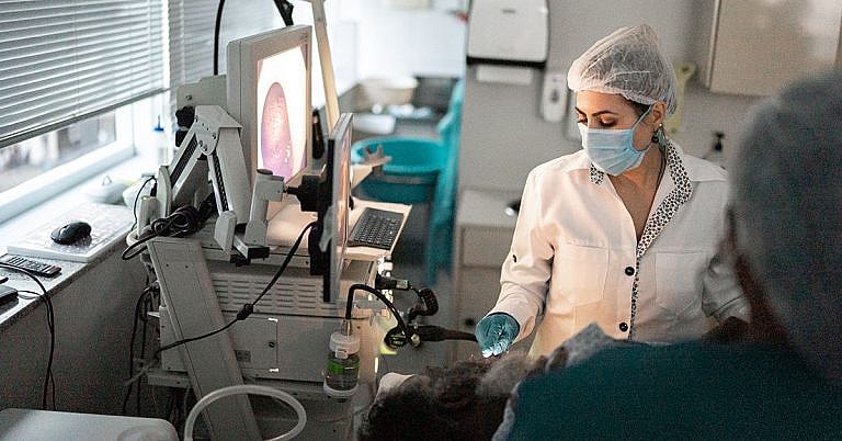
To take photographs, the patient is asked to lie down on an exam table. Electrodes are then placed on the chest, enabling the cardiovascular perfusionist to monitor the patient’s heart rate during the test. With the patient’s arms raised above his head, the practitioner will position a number of cameras around the patient’s heart to take a series of photographs for about 15 minutes. Patients are advised to stay still during this part of the procedure to ensure the clarity and accuracy of the photographs.
The second part of the test involves giving the patient dobutamine, a drug that simulates the effects of exercise on the heart, for about 12 minutes. When the patient’s heart rate reaches a certain level, another injection of the same radioactive tracer is given, and the distribution of the tracer in the heart is once again captured through a set of photographs. This will take another 20 minutes, after which the patient is given a short break.
After the break, another set of photographs will be taken. This part of the test will take another 30 minutes. All in all, the entire procedure takes approximately three hours, although in some cases, it may last for as long as 4 and ½ hours.
Possible Risks and Complications
Certain precautions have to be taken to ensure patient safety during the procedure. This is because the dobutamine drug may cause some potentially harmful side effects, such as:
Heart flutter
Chest pain or angina
Cardiac arrhythmias
Premature atrial or ventricular contractions
Supraventricular and ventricular tachycardias
Hypotension
Flushing
However, the efficacy of the drug only lasts for a short time, so side effects, if any, usually pass quickly or once the infusion of the drug is stopped. As such, the test has been confirmed safe for use even in elderly and heart transplant patients, and those suffering from left ventricular dysfunction.
Other non-cardiac side effects may also occur, but these are generally uncommon and are well-tolerated by patients. These include:
Mild nausea
Headache
Chills
Additionally, there is some degree of risk due to the use of the radioactive substance, which releases a small amount of radiation. However, this amount is very small and is well within the safety guidelines.
To prevent complications, patients are advised to refrain from taking products that contain caffeine the day before the test. Beta-blocker medications should also be avoided 48 hours prior to the scan. To ensure that no dangerous drug interactions occur, patients are advised to provide their attending physician with a complete list of all medications and supplements that they are taking.
References:
Geleijnse M., Elhendy A., et al. “Dobutamine stress myocardial perfusion imaging.” J Am Coll Cardiol. 2000;36(7):2017-2027. http://content.onlinejacc.org/article.aspx?articleid=1126864
Abdou e., Bax J., Poldermans D. “Dobutamine Stress Myocardial Perfusion Imaging in Coronary Artery Disease.” The Journal of Nuclear Medicine. http://jnm.snmjournals.org/content/43/12/1634.full
/trp_language]
[trp_language language=”ar”][wp_show_posts id=””][/trp_language]
[trp_language language=”fr_FR”][wp_show_posts id=””][/trp_language]
**What is Dobutamine Stress Myocardial Perfusion Imaging?**
**Overview**
Dobutamine stress myocardial perfusion imaging (DSE MPI) is an advanced cardiac imaging technique that combines dobutamine, a medication that stimulates the heart muscle, with myocardial perfusion imaging. It helps assess blood flow to the heart muscle (myocardium) during stress and rest conditions.
**Benefits of DSE MPI**
* Detects coronary artery disease (CAD), even in patients with no symptoms.
* Evaluates the significance of coronary artery blockages.
* Determines the effectiveness of coronary interventions (e.g., bypass surgery, stenting).
* Tracks disease progression and monitors therapy response.
**Expected Results**
DSE MPI provides a detailed map of blood flow to the heart muscle, showing areas of:
* **Normal perfusion:** No abnormalities in blood flow.
* **Diminished perfusion:** Reduced blood flow to a particular area, typically indicating a CAD blockage.
* **Reversible perfusion defect:** Diminished perfusion during stress that improves during rest, suggesting viable heart muscle downstream of the blockage.
* **Fixed perfusion defect:** Diminished perfusion in both stress and rest conditions, indicating irreversible heart muscle damage.
**Procedure**
* Patient receives an intravenous line for contrast dye administration.
* Patient lies on a scanning table and electrodes are attached to their chest.
* Dobutamine is administered through the IV.
* Multiple images are taken with a special camera that detects the radioactive tracer.
* Rest images are taken later, generally after 24 hours.
**Risks and Limitations**
DSE MPI is generally safe, but potential risks include:
* Allergic reaction to contrast dye.
* Arrhythmias (abnormal heart rhythms).
* Chest discomfort or pain.
Limitations of DSE MPI include:
* May not be suitable for patients with severe CAD or those who cannot tolerate dobutamine.
* Results can be influenced by medications and other factors.
**Conclusion**
Dobutamine stress myocardial perfusion imaging is a valuable tool for evaluating heart muscle blood flow and detecting coronary artery disease. By understanding the benefits, expected results, and potential limitations of DSE MPI, patients can make informed decisions about their cardiac care.


Thanks for your helpful post.
## Overview of Dobutamine Stress Myocardial Perfusion Imaging