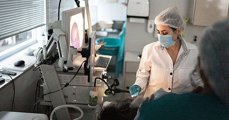
What is Ventricular Septal Defect Closure: Overview, Benefits, and Expected Results
The new product is a great addition to our lineup.
Our latest product is an exciting addition to our already impressive lineup! With its innovative features and sleek design, it's sure to be a hit with customers. Don't miss out on this amazing opportunity to upgrade your life!
What is Ventricular Septal Defect Closure?
Ventricular Septal Defect (VSD) Closure is a medical procedure used to repair a hole in the heart known as ventricular septal defect (VSD). This defect affects the muscle that divides the left and right ventricles, and can be present at birth or develop over time. VSD Closure is a minimally invasive surgical procedure designed to close the defect and repair the heart’s functioning.
Overview
VSD is a congenital heart defect that affects up to one in every 1000 live births. It occurs when a hole develops between the two lower chambers of the heart, known as ventricles. This allows oxygen-rich blood to leak into the oxygen-poor lower chamber, leading to an imbalance in the oxygen levels resulting in various related complications.
Patients may not experience any symptoms or may have only mild symptoms such as breathing difficulty, poor weight gain and fatigue. More serious cases may cause an arrhythmia (irregular heart beat) and heart failure, which is why prompt treatment and repair are essential.
Methods of Closure
VSD Closure is usually performed using a minimally invasive procedure known as transcatheter device closure. This involves a surgical opening in the chest through which a catheter is passed, directing the closing device to the affected area. The device is then used to push the edges of the defect together. This is the most common procedure and is generally used for patients with small- to medium-sized defects.
A surgical closure may be recommended for patients with large-sized defects. This involves the opening of the chest to allow access to the heart, and will require a general anesthetic. Surgical repair is more complex and carries a higher risk of complications, yet some may be the better option for larger defects.
Benefits
Improved Blood Flow
By closing the defect, VSD Closure repairs the flow of blood throughout the heart, which reduces stress on the heart and increases oxygen levels in the body. This improved blood flow helps to reduce the risk of, or treat, associated complications such as arrhythmia and heart failure.
Minimally Invasive
VSD Closure is a minimally invasive procedure. The patient is usually kept awake throughout, and no general anesthetic is required. The device used to close the hole is much smaller than conventional surgical instruments, meaning the procedure can be completed with fewer and smaller incisions. This reduces recovery time and reduces the risk of associated complications.
Improved Quality of Life
VSD Closure restores the heart’s normal functioning, improving the patient’s quality of life. VSD Closure helps to improve breathing quality, reduce fatigue and improve overall energy levels. In more serious cases, this improved quality of life can often be life-saving.
Expected Results
The expected results of VSD Closure will depend on the patient’s individual case. Generally, the procedure is performed to reduce the risk of developing complications or to reverse existing damage.
For patients with an arrhythmia, VSD Closure helps to reduce abnormal heart beat and improve circulation. It can also reduce the risk of developing future arrhythmias and improve quality of life.
For patients with heart failure, VSD Closure helps to improve the heart’s functioning and can often reverse existing damage. It can help to reduce symptoms such as shortness of breath, fatigue, and poor weight gain.
Long-term results may depend on the patient’s lifestyle and medical history. Patients are advised to follow their doctor’s advice for optimal results.
FAQs About VSD Closure
What is Ventricular Septal Defect (VSD) Closure?
Ventricular Septal Defect (VSD) Closure is a medical procedure used to repair a hole in the heart known as ventricular septal defect (VSD). This defect affects the muscle that divides the left and right ventricles, and can be present at birth or develop over time. VSD Closure is a minimally invasive surgical procedure designed to close the defect and repair the heart’s functioning.
Are there any risks associated with VSD Closure?
VSD Closure is generally very safe. However, there is a risk of complications associated with both the transcatheter and surgical procedures. These can include damage to the heart muscle, an infection, or a blockage of the heart’s arteries.
What is the recovery process like?
The recovery process for VSD Closure is typically very fast. Patients may be able to leave the hospital the same day or the day after the procedure. It is important to follow the doctor’s recommendations for recovery, such as taking medications as prescribed and not participating in strenuous activities for a few weeks.
How long do the results of VSD Closure typically last?
The results of VSD Closure are generally long-lasting. The defect should remain closed and the patient’s condition should remain stable over time. It is important to follow up with the doctor regularly for check-ups to make sure the heart remains in good condition.
Conclusion
VSD Closure is a minimally invasive procedure used to repair a hole in the heart known as VSD. The procedure helps to restore normal heart functioning and can improve the patient’s quality of life. It is generally very safe and the recovery process is typically short. By following the doctor’s advice, the patient’s condition should remain stable and the results of VSD Closure should be long-lasting.
Definition and Overview
A ventricular septal defect (VSD) is the most common congenital heart disease, occurring in 2 per 1000 live births. It is a hole in the ventricular septum, which separates the left and right ventricles. A VSD may be an isolated defect or it may be associated with more complex lesions, such as tetralogy of Fallot and atrioventricular canal defects. In most patients, a VSD spontaneously closes. However, some patients will require ventricular septal defect closure surgery to alleviate the symptoms and prevent the development of irreversible heart failure.
VSDs may also develop in adult patients and are usually caused by a traumatic or an ischemic (heart attack) etiology. These VSDs must be closed surgically.
Who Should Undergo and Expected Results
A ventricular septal defect is a left-to-right shunt, which means that oxygenated blood coming from the left ventricle is pumped to the right side and re-circulated to the pulmonary bed. This causes an increase in pulmonary flow, which results in increased workload for both ventricles, as well as an increased risk of recurrent infections of the respiratory tract.
Patients with VSDs typically present with symptoms of congestive heart failure, such as difficulty of breathing and failure to thrive. A harsh murmur is heard upon auscultation, and further examination may reveal the enlargement of the left ventricle and left atrium. Approximately 30% of patients with ventricular septal defects develop failure symptoms within the first year of life, necessitating ventricular septal defect closure.
Persistent shunting across the VSD also results in changes in the pulmonary vasculature. Through time, this can lead to pulmonary hypertension, a serious complication of an unrepaired VSD. This condition requires surgical intervention before changes become irreversible.
There are several kinds of VSD, depending on the location of the defect on the septum. The perimembranous kind is the most common. Inlet type and outlet type VSDs are not likely to spontaneously close, and should be repaired once diagnosed. Aortic regurgitation or insufficiency can accompany VSDs, especially the outlet type, due to prolapse of the aortic valve into the defect. Surgery is likewise indicated in VSD patients with progressive aortic insufficiency.
The closure of an isolated VSD is generally associated with good outcomes. The mortality rate is very low, approaching 0% in most institutions, even in extremely small infants. Good candidates who undergo the procedure within the first two years of life are generally able to achieve full functionality, and life expectancy is normal or near normal.
How is the Procedure Performed?
Ventricular septal defect closure is performed under cardiopulmonary bypass. The majority of isolated VSDs are approached via an incision in the right atrium. The tricuspid valve, located between the right atrium and the right ventricle, is retracted to access the ventricular septum. A patch, which may be made of polytetrafluoroethylene (PTFE or Gore-Tex), Dacron or pericardium, is fashioned to the size of the defect. The patch is then sutured to the ventricular septum and the tricuspid annulus. Caution should be exercised when suturing the patch, making sure that vital structures, such as the conduction system and the aortic valve, are not injured or affected. If there are other associated conditions, such as a patent ductus arteriosus or aortic insufficiency, they are also addressed during the same procedure. Other kinds of VSDs (outlet type) and associated defects may have to be approached through additional incisions.
Patients are typically extubated within the first 24 hours after surgery. Temporary pacing wires may be inserted during the procedure and are useful in cases of postoperative arrhythmias.
Possible Risks and Complications
Patients undergoing VSD closure are at risk of developing complications associated with the use of cardiopulmonary bypass. These include bleeding, kidney failure and pneumonia, among others.
Aside from these, there are also complications associated with the procedure itself. Injury to vital structures near the defect, such as the tricuspid valve and aortic valve, may occur during the placement of sutures.
Since the conduction system of the heart travels through the ventricular septum, there is a risk for the development of rhythm abnormalities. Right bundle branch blocks occur commonly but are usually tolerated well by the patient. Complete heart block occurs in approximately 1-3% of patients and this may necessitate the insertion of a permanent pacemaker.
Very young patients subjected to VSD closure are at risk for residual VSDs. This usually occurs because of suture dehiscence through the friable cardiac muscle in small infants. In most patients, these can be observed and will eventually close spontaneously. However, if the leak is large and causes hemodynamic problems, reoperation should be done.
In older patients, pulmonary hypertension is a dreaded complication after VSD closure. Sedation, hyperventilation, and the administration of nitric oxide are useful techniques in the management of postoperative pulmonary hypertensive crisis. These patients usually have decreased exercise tolerance and are at risk of premature death in the long-term.
References:
Muller WH, Damman JF. The treatment of certain congenital malformations of the heart by the creation of pulmonic stenosis to reduce pulmonary hypertension and excessive pulmonary blood flow; a preliminary report. Surg Gynecol Obstet. 1952 Aug. 95(2):213-9.
Quansheng X, Silin P, Zhongyun Z, Youbao R, Shengde L, Qian C, et al. Minimally invasive perventricular device closure of an isolated perimembranous ventricular septal defect with a newly designed delivery system: preliminary experience. J Thorac Cardiovasc Surg. 2009 Mar. 137(3):556-9.
/trp_language]
[trp_language language=”ar”][wp_show_posts id=””][/trp_language]
[trp_language language=”fr_FR”][wp_show_posts id=””][/trp_language]


Super helpful information! #knowledgeispower #medicine
Great article! Everything is explained clearly.