What is Release of Ureter: Overview, Benefits, and Expected Results
The new product is a great addition to our lineup.
Our latest product is an exciting addition to our already impressive lineup! With its innovative features and sleek design, it's sure to be a hit with customers. Don't miss out on this amazing opportunity to upgrade your life!
What is Release of Ureter? Overview, Benefits, and Expected Results
Ureter release is a surgical procedure used to treat vesicoureteral reflux, a disorder that impacts the flow of urine from the bladder to the kidneys. This sequence of events describes what happens during the procedure, the benefits, and expected results.
What is Vesicoureteral Reflux?
Vesicoureteral Reflux (VUR) is a condition when urine from the bladder flows back into the ureters and kidneys. It affects up to 30% of children diagnosed and can cause mild to severe urinary tract infections (UTIs) and damage to the kidneys. Children with VUR experience painful urination, bed-wetting, dark and bloody urine, as well as abdominal and flank pain or discomfort. This condition is usually brought to doctors’ attention when UTIs are diagnosed.
What is Ureter Release Surgery?
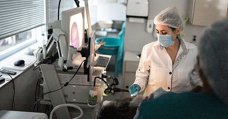
Ureter release surgery is a minimally-invasive procedure used to treat VUR that involves detaching part of the bladder from the ureters, thus allowing urine to flow unhindered. It may be done in either a laparoscopic or open technique, where a unique combination of high-resolution imaging and specialized instruments are used to perform the procedure safely and effectively.
During typical laparoscopic ureter release surgery, a small incision is made in the abdomen or groin area. A miniature camera attached to a specialized instrument is then inserted into the body to identify the ureters and nephrostomy tube. The surgeon then uses the instrument to separate the bladder from the ureters and carefully reposition the bladder into its normal location.
Benefits of Ureter Release Surgery
Ureter release surgery offers several benefits to patients with VUR. These include:
- Minimally-invasive: Ureter release surgery is minimally-invasive, which means patients experience less pain and trauma during the procedure, as well as reduced recovery times. Additionally, since laparoscopic ureter release uses cameras and specialized instruments, surgeons can easily correct VUR, even in difficult-to-reach areas of the body.
- Relief of symptoms: Ureter release surgery can decrease the frequency and severity of symptoms associated with VUR, such as painful urination, bed wetting, and dark or bloody urine.
- Protection of kidneys from damage: Ureter release surgery is effective at preventing further damage to the kidneys from recurring UTIs.
Expected Results
Ureter release surgery is effective at improving the flow of urine from the bladder to the kidneys. Patients who undergo the procedure can expect to experience fewer UTIs and symptoms associated with VUR, as well as improved kidney and bladder health.
For the first few weeks after the surgery, patients may experience light to moderate discomfort, including swelling and soreness in the hips, groin, and abdomen. Pain medications are typically prescribed to minimize any pain or discomfort. Most patients regain their normal activity levels within a few weeks after surgery.
It is important to follow instructions given by your doctor after the procedure, including taking all prescribed medications, avoiding heavy lifting and strenuous activities, and attending follow-up appointments. Depending on the patient’s age and the severity of the VUR, additional monitoring may be necessary, such as using imaging techniques to track the progress of the condition.
Conclusion
Ureter release surgery is an effective procedure to treat vesicoureteral reflux. The procedure is minimally-invasive, can provide relief of symptoms, and protect the kidneys from further damage. Patients undergoing the procedure can expect to experience fewer UTIs, and improved kidney and bladder health. It is important to follow instructions given by the doctor and attend all follow-up appointments.
Definition and Overview
The release of ureter is a procedure used to treat ureteral obstructions and strictures. By releasing and draining the ureters in such cases, the procedure allows the kidneys to function normally.
The ureter is a small thin tube that connects the kidney to the bladder. If it is blocked or obstructed, it has to be released so it can resume transporting urine to the bladder and prevent further complications.
Who Should Undergo and Expected Results
The procedure to release the ureter is necessary for patients who suffer from ureteral obstruction, which may be caused by:
Congenital/developmental problem – The ureteropelvic junction (UPJ), which is located in the upper abdomen, connects the kidney and the ureter to each other. Certain congenital or developmental abnormalities can affect how the kidney pushes urine towards the UPJ and into the ureter (a process called peristalsis).
Ureteral strictures (scarring) – The ureter can sometimes develop scarring due to previous surgery, which can cause kidney blockage.
Retroperitoneal fibrosis – Refers to the inflammation of abdominal organs around the ureter.
Urethritis – Refers to the inflammation of the urethra.
Kidney stones – A stone in the kidney may cause kidney obstruction and produce the same complications as urethral obstruction.
Cancer – Some cancerous tumours in the abdomen may push through or compress the ureter. These include cervical, ovarian, uterine, colon, prostate, and bladder cancers.
Ureteral stricture, which is one of the most common reasons why the release of ureter is performed, is a common complication of some surgical procedures that are performed on structures that are close to the ureter. The most commonly associated surgeries include:
Kidney stone removal surgery
Abdominal vascular surgery, or surgery involving the arteries in the abdomen
Obstetric or gynaecologic surgery, such as the removal of the ovaries or uterus – The link between gynaecologic surgery and urethral strictures is due to the close proximity of the ureters to the arteries of the uterus and ovaries. If the ureter injury is only detected after the operation is complete, it may not be treated immediately due to the inflammation that normally occurs after a surgical procedure. In such cases, doctors have to wait between 6 and 12 weeks before the ureter can be surgically released. To facilitate proper urination, a nephrostomy tube, which bypasses the stricture, is used to drain out the urine from the kidney.
After the ureter has been released, patients can expect relief from abdominal pain as the kidney, ureter, and bladder start working properly again.
How is the Procedure Performed?
The manner in which the procedure is performed differs depending on the cause of the problem and the location of the blockage or stricture.
If the problem is caused by blockage or the compression of the ureter, the ureter is simply drained using a ureteral stent or nephrostomy tube.
Ureteral stent – Also called a ureteral double-J stent, this is an internal tube that connects the kidney to the bladder to take over the function of the ureter. If the stent is used for kidney stones, it is also called a kidney stone stent. The kidney stent bypasses the part where the stone is located to allow urine to pass into the bladder.
Nephrostomy tube – An external tube that drains the contents of the kidney into a bag.
The ureteral or kidney stent and nephrostomy tube can be eventually removed, such as when the cause of the obstruction has been treated. An example is when a cancerous tumour has been surgically removed or shrunk by other treatments.
In some cases, the ureter has to be repaired or reconstructed after it was released. This is common among patients who suffer from urethral strictures or scarring that has caused permanent damage to some parts of the ureter.
If the ureter is damaged or scarred due to a previous abdominal surgery, the type of procedure used to release it depends on the location of the damage or scarring. If it is located in the lower part of the ureter and is closer to the bladder, the scarred part of the ureter is simply removed and the ureter is reconnected to the bladder. This procedure is called neo-cystotomy.
If the urethral stricture is too long, the ureter may need to be reconstructed once the scarred part is removed. Doctors can use either a flap taken from the bladder (Boari flap) or small bowel (ileal ureter) as a substitute.
Possible Risks and Complications
The release of the ureter is a generally safe procedure. However, patients still face a low risk of certain complications. These include:
Dislocation of the ureteral stent
Infection
Encrustation leading to blockage (also known as encrusted stents) – Factors that increase a person’s risk of encrusted stents include pregnancy, chronic renal failure, chemotherapy, and congenital abnormalities
Increased urination urgency and frequency
Bloody urine
Urine leakage
Kidney pain
Bladder pain
Pain in the kidneys during urination
Irritated urethra
Some of these complications are temporary and may disappear when the stent is removed. More recently, surgeons have begun using stents with heparin coatings. These have been proven to help reduce the risk of infection and encrustation.
References:
Ahallal Y, Khallouk A, El Fassi MJ, Farih MH. “Risk Factor Analysis and Management of Ureteral Double-J Stent Complications.” Rev Urol. 2010 Spring-Summer; 12(2-3): e147-e151. https://www.ncbi.nlm.nih.gov/pmc/articles/PMC2931292/
“Ureteral obstructions and strictures.” University of Utah. https://healthcare.utah.edu/urology/conditions/ureteral/
Breyer BN. “Ureteral stricture.” Medscape. http://emedicine.medscape.com/article/442469-overview
Vasavada SP. “Ureteral injury during gynecologic surgery.” http://emedicine.medscape.com/article/454617-overview
Santucci RA. “Ureteral trauma treatment and management.” http://emedicine.medscape.com/article/440933-treatment
Dyer RB, Chen MY, Zagoria RJ, Regan JD, Hood CG, Kavanagh PV. “Complications of ureteral stent placement.” RadioGraphics 2002; 22:1005-1022. http://pubs.rsna.org/doi/pdf/10.1148/radiographics.22.5.g02se081005
/trp_language]
[trp_language language=”ar”][wp_show_posts id=””][/trp_language]
[trp_language language=”fr_FR”][wp_show_posts id=””][/trp_language]


Interesting! #ureter
Great reading! #medical
Very helpful #ureterrelease