4D Scan
Definition & Overview
A 4D scan is a prenatal ultrasound that incorporates time as the 4th dimension allowing it to capture not just still images but also a video of a baby inside a womb. It can be performed for medical reasons, such as when birth defects that do not show up on a standard 2D and 3D scans are suspected or as an elective procedure. However, experts do not advocate having 4D scans purely for souvenir photos and videos as it can expose the unborn child to more ultrasound that is medically necessary.
Who Should Undergo and Expected Results
All pregnant women can elect to undergo a 4D ultrasound scan. However, it is only typically recommended by doctors if some birth defects, congenital anomalies, or medical conditions that affect the mother are suspected. These include:
- Neural tube defects
- Foetal aneuploidy
- Cleft palate, which is not detected by a standard 2D scan
- Uterine fibroids
- Subchorionic hemorrhage
- Ovarian mass
The standard prenatal imaging scan used nowadays is 2D or two-dimensional, which captures images of a foetus’ internal organs and bones. A 3D scan, on the other hand, is another optional scan that can produce images of the baby from the surface, but cannot capture movements like a 4D scan can. This makes the latter the most advanced ultrasound technology currently available.
Like other ultrasound scans, a 4D scan can be used to:
- Confirm normal foetal development
- Confirm the patient’s due date and the baby’s gestational age
- Detect or confirm multiple pregnancies
- Check for other issues that may affect the pregnancy
- Check the position of the placenta
In order to obtain accurate results, 4D ultrasound should be performed between 26th and 30th weeks of pregnancy. Around this time, the baby will have sufficient subcutaneous fat, so the surface of the skin will be more accurately captured during the scan.
How is the Procedure Performed?
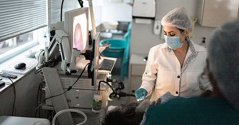
For the procedure, the patient lies down on an exam table and a gel-like substance, which is responsible for carrying the sound waves, is applied to her abdomen. A probe is then placed against the abdomen and moved around to produce images that instantly show up on a computer monitor. The images are then examined to check for anything that may seem unusual. The procedure takes 15-20 minutes and is generally painless and comfortable. It does not require any type of anaesthesia.
Possible Risks and Complications
Due to the use of ultrasound energy, experts advice doctors and expectant parents alike to use 4D scans minimally and with caution. Although there are studies that show that the procedure is as safe as standard 2D scans and that the levels of ultrasound exposure of both scans are the same, some argue that 4D ultrasound exposes the unborn child to more ultrasound that is medically necessary. For this reason, some doctors only allow 4D scans when there is a medical reason to perform them.
References:
Tomasovic S., Predojevic M. “4D Ultrasound – Medical Devices for Recent Advances on the Etiology of Cerebral Palsy.” Acta Inform Med. 2011 Dec; 19(4):228-234. http://www.ncbi.nlm.nih.gov/pmc/articles/PMC3564175/
Kurjak A., Predojevic M., Kadic A. “Fetal Behavior in 4D Ultrasound in the Progress of Perinatal Medicine.” Journal of Health and Medical Informatics. http://www.omicsonline.org/fetal-behavior-in-4d-ultrasound-in-the-progress-of-perinatal-medicine-2157-7420.S11-006.php?aid=13161
/trp_language]
[trp_language language=”ar”][wp_show_posts id=””][/trp_language]
[trp_language language=”fr_FR”][wp_show_posts id=””][/trp_language]
**4D Scan: A Comprehensive Guide**
**Q: What is a 4D scan?**
**A:** A 4D scan, also known as a 4D ultrasound, is a type of prenatal imaging that combines 3D ultrasound with real-time imaging to create a live, moving image of the developing baby.
**Q: When is a 4D scan typically performed?**
**A:** 4D scans are typically performed between 24 and 32 weeks of gestation, when the baby’s facial features and movements are most visible.
**Q: What benefits does a 4D scan offer?**
**A:** 4D scans offer many benefits, including:
* Providing a more comprehensive view of the baby’s anatomy
* Detecting potential health issues more accurately
* Allowing parents to see and interact with their baby before birth
* Capturing special moments, such as the baby’s first movements
**Q: Are there any risks associated with 4D scans?**
**A:** 4D scans are generally considered safe for both the mother and the baby. However, it’s important to note that the additional time spent in the ultrasound room may increase the exposure to ultrasound energy.
**Q: How should I prepare for a 4D scan?**
**A:** No special preparation is required for a 4D scan. Simply drink plenty of fluids beforehand to have a full bladder, which helps to provide better images.
**Q: What should I expect during a 4D scan?**
**A:** During a 4D scan, you will lie on a padded table and a warm ultrasound gel will be applied to your abdomen. A wand-like device emits sound waves that create images of the baby on a monitor. The images can be captured and printed as 3D or 4D photos or videos.
**Q: Can I get a 4D scan at any hospital or clinic?**
**A:** Not all hospitals or clinics offer 4D scans. It’s best to contact your healthcare provider or local imaging center to inquire about availability and costs.


I can’t wait to see what my baby looks like! I’ve heard that 4D scans are amazing.
kariztecaylor:
My baby is the cutest little thing! I just love looking at his 4D scan.
laurengjones:
I’m so glad I got a 4D scan. It was such a special experience to see my baby moving around and yawning.
I’m so excited to get a 4D scan of my baby! I’ve heard that they’re amazing.