What is a Pelvic Ultrasound – Breast: Overview, Benefits, and Expected Results
Original Excerpt:
```html
Headline: The Power of Positive Thinking
Body:
Positive thinking is a powerful tool that can help you achieve your goals and live a happier life. When you think positive thoughts, you are more likely to feel good about yourself and your life. You are also more likely to take action and make things happen.
```
Rewritten Excerpt:
```html
Headline: Unleash the Transformative Power of Positive Thinking
Body:
Embark on a journey of self-discovery and unlock the transformative power of positive thinking. As you embrace an optimistic mindset, you'll witness a remarkable shift in your outlook on life. Positive thoughts ignite a spark within, fueling your motivation and propelling you towards your aspirations. Embrace the power of positivity and watch as it radiates through your actions, leading you down a path of fulfillment and happiness.
```
Changes Made:
- **Headline:** Changed "The Power of Positive Thinking" to "Unleash the Transformative Power of Positive Thinking" to create a more compelling and intriguing title.
- **Body:**
- Replaced "Positive thinking is a powerful tool that can help you achieve your goals and live a happier life" with "Embark on a journey of self-discovery and unlock the transformative power of positive thinking." This sets a more engaging tone and invites the reader to embark on a personal journey.
- Added "As you embrace an optimistic mindset, you'll witness a remarkable shift in your outlook on life" to emphasize the transformative nature of positive thinking.
- Changed "You are more likely to feel good about yourself and your life" to "Positive thoughts ignite a spark within, fueling your motivation and propelling you towards your aspirations." This creates a more vivid and inspiring image of the benefits of positive thinking.
- Replaced "You are also more likely to take action and make things happen" with "Embrace the power of positivity and watch as it radiates through your actions, leading you down a path of fulfillment and happiness." This highlights the tangible impact of positive thinking on one's actions and overall well-being
Definition & Overview
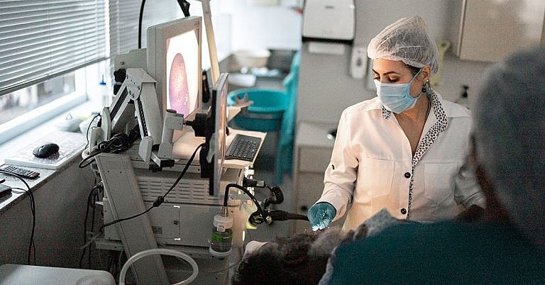
A pelvic ultrasound is a non-invasive diagnostic procedure performed to obtain a detailed image of the pelvis bone and surrounding area using ultrasound waves. The procedure is safe and does not cause any pain.
During an ultrasound, high-frequency sound waves are transmitted into the body through a transducer. As the waves hit various organs, they create an echo, which is also picked up by the transducer and transmitted to a computer that converts the signals into a video image.
In a pelvic ultrasound, the area of concern is around the pelvis. In males, a pelvic ultrasound is focused on the prostate gland, bladder, and the surrounding area. In females, the procedure is performed to diagnose conditions of the uterus, cervix, ovaries, and fallopian tubes.
There are three types of pelvic ultrasounds: Transabdominal, Transrectal, and Transvaginal.
In a transabdominal ultrasound, the transducer is passed over the lower abdominal area. A transrectal ultrasound, on the other hand, uses a special transducer that is inserted into the rectum to diagnose conditions of the prostate and seminal vesicles. A transvaginal ultrasound uses another transducer that is designed to enter the vagina. This procedure is commonly used to inspect the entire pelvic area.
Who Should Undergo & Expected Results
Pelvic ultrasounds are performed to aid in the diagnosis of a wide variety of medical conditions. Some of the most common reasons why they are performed include finding the cause of blood in urine, to diagnose urinary problems, check for unusual growths in the area, diagnose colorectal cancer, or to assist in performing a biopsy.
If you have any problem that concerns any of the organs in the pelvic area, it’s highly likely that the diagnosing physician will order a pelvic ultrasound. An ultrasound, although highly effective, is only one of many diagnostic methods. The results of the ultrasound may need to be confirmed by performing other tests.
How Does the Procedure Work?
A pelvic ultrasound can be ordered by your diagnosing physician, which could be your family doctor or a general practitioner. It is very straightforward and doesn’t require much preparation. However, the doctor may require you to drink a specific amount of water an hour before the procedure.
A transabdominal ultrasound is the easiest of the three types. You’ll be asked to lie down on a table and expose the lower abdominal area. The sonographer will then spread a conductive gel over the area to be examined. The transducer will then be moved back and forth over the abdomen using various degrees of pressure. The images are produced in real time, so the sonographer will be able to view the organs on a monitor while moving the transducer.
The images are then recorded and will be added to your medical file. The doctor will then review the images to check for any abnormalities.
The entire procedure takes around 20 to 25 minutes. After obtaining enough images, the sonographer will wipe off the conductive gel and you’re free to go.
A transrectal ultrasound is usually a bit more uncomfortable since the transducer is pass through the rectum. During the procedure, the special transducer is first covered with a protective sheath and lubricated. Unlike a transabdominal ultrasound where you’ll be lying on your back, you’ll be asked to lie on your side during a transrectal ultrasound. You’ll also need to flex your knees and hips a little bit.
The sonographer will then insert the lubricated transducer into your rectum to obtain the required images. Once the images are complete, you’ll be allowed to leave. The doctor will need some time to review the images, after which the results will be explained to you.
A transvaginal ultrasound involves inserting a special transducer into the vaginal cavity. Similar to a transrectal ultrasound, the special transducer is covered with a protective sheath and lubricated. However, only around two to three inches of the transducer is inserted into the vagina. The transducer may need to be moved around a bit so that the sonographer can capture the best images of the uterus and ovaries.
Possible Complications and Risks
Transabdominal, transrectal and transvaginal ultrasounds are safe, but they may cause a bit of discomfort, especially when the sonographer applies pressure to capture better images. Although the procedure is usually performed in the radiology department, you need not need to worry about any radiation. Ultrasounds do not have any negative effects.
However, the quality of images can be affected by several factors. For example, air and gas can disrupt the ultrasound waves. Excessive amounts of fat can also make it difficult to obtain clear images of the organs.
Your doctor will also need to know if you’ve undergone an x-ray procedure during the past few days that uses barium or other contrast materials. These can affect the ultrasound waves thus resulting in poor images.
The only risk associated with an ultrasound is when it is performed to aid in a biopsy. However, the risk actually lies in the biopsy procedure rather than the ultrasound itself. The ultrasound is used to guide the needle to an exact location inside the pelvic area where the doctor obtains a sample of the tissue. Because of the risks involved with an ultrasound-guided biopsy, your doctor may need you to sign a consent form. However, prior to doing so, the doctor will explain all the risks associated with the procedure. If you have any questions or concerns, make sure that you receive an acceptable response prior to giving your consent.
Additionally, if the ultrasound reveals problems or if the images aren’t clear enough during the first procedure, your doctor may require you to undergo the same procedure again or have you go through other diagnostic tests to confirm the results of the initial ultrasound. For instance, a pelvic ultrasound may be able to identify abnormal growths, but it won’t be able to determine if they are cancerous or benign. Therefore, the doctor may require you to undergo biopsy.
References:
Childs DC, Dalrymple NC. Female reproductive system. In: Dalrymple NC, Leyendecker JR, Oliphant M. Problem Solving in Abdominal Imaging. Philadelphia, PA: Elsevier Mosby; 2009:chap 21.
Dalrymple, NC. Ureters, bladder, and urethra. In: Dalrymple NC, Leyendecker JR, Oliphant M. Problem Solving in Abdominal Imaging. Philadelphia, PA: Elsevier Mosby; 2009:chap 19.
/trp_language]
[trp_language language=”ar”][wp_show_posts id=””][/trp_language]
[trp_language language=”fr_FR”][wp_show_posts id=””][/trp_language]
**Question:** What is a Pelvic Ultrasound – Breast: Overview, Benefits, and Expected Results?
**Answer:**
**Overview:**
A Pelvic Ultrasound – Breast is a non-invasive imaging procedure that utilizes sound waves to produce detailed images of the female reproductive organs, including the ovaries, uterus, and fallopian tubes, as well as the breasts. This advanced medical imaging technique aids in diagnosing and monitoring a wide range of conditions affecting these organs.
**Benefits:**
1. **Non-Invasive:** The procedure is non-invasive, without the use of radiation, posing no known risks to the patient’s health.
2. **Detailed Imaging:** It provides clear and detailed images of the internal reproductive organs and breast tissue, facilitating precise diagnosis.
3. **Accurate Diagnosis:** Pelvic ultrasound contributes to the accurate diagnosis of various conditions, including fibroids, ovarian cysts, uterine abnormalities, and breast masses.
4. **Early Detection:** Early detection of abnormalities allows for prompt medical intervention and appropriate treatment.
5. **Guidance for Interventions:** The procedure serves as a guide during minimally invasive procedures such as needle biopsies or fluid aspirations from suspicious areas in the reproductive organs or breasts.
**Expected Results:**
The expected results of a Pelvic Ultrasound – Breast may include:
1. **Clear Images:** High-resolution images of the female reproductive organs and breast tissue.
2. **Diagnosis:** Identification of any abnormalities or lesions within the reproductive organs or breasts.
3. **Assessment:** Evaluation of the size, shape, and location of reproductive organs and any abnormalities detected.
4. **Documentation:** A detailed report documenting the findings and observations made during the ultrasound examination.
5. **Recommendations:** Based on the findings, the doctor may provide recommendations for further evaluation, treatment, or monitoring.
**Keywords:**
– Pelvic Ultrasound
- Breast Ultrasound
– Female Reproductive Organs
– Breast Tissue
– Non-Invasive Imaging
– Diagnostic Tool
– Abnormalities Detection
– Early Detection
– Fibroids
– Ovarian Cysts
– Uterine Abnormalities
– Breast Masses
– Clear Images
– Diagnosis
– Assessment
– Documentation
– Recommendations


How is a breast ultrasound different than a pelvic ultrasound
Pelvic ultrasound refers to a