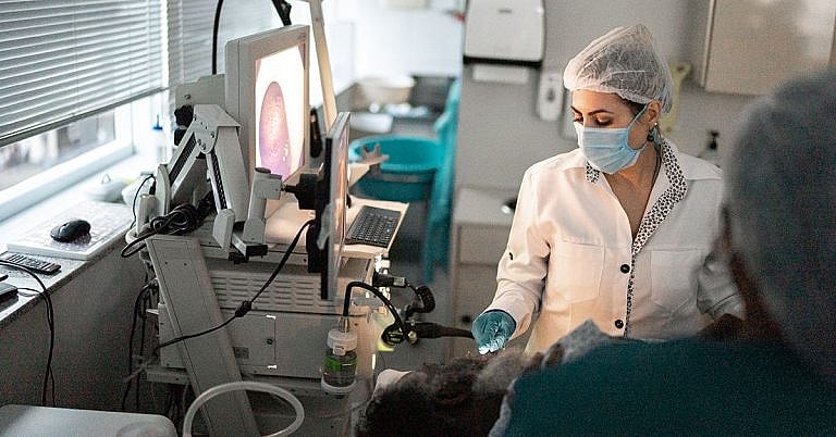
What is Cerebrospinal Fluid Flow Imaging or Cisternogram Scan : Overview, Benefits, and Expected Results
Definition & Overview
Cisternogram scan is a cerebrospinal fluid flow imaging technique. It is one of the many methods used to evaluate the flow of cerebrospinal fluid (CSF). The purpose of the procedure is to detect a cerebrospinal fluid leak. The test can determine exactly where the leakage is located.
A cisternogram scan can be performed using a contrast dye material (computed tomography) or with nuclear medicine using a radionuclide material. Due to the limitations of both methods, they are sometimes combined to produce the best anatomic resolution and the highest sensitivity.
Who Should Undergo and Expected Results
Cisternogram scan is beneficial for:
- Patients suffering from normal pressure hydrocephalus
- Patients suspected of having abnormal spinal fluid flow
- Patients who have suffered from or are suspected of having a cerebrospinal fluid leak. The leak is caused by a tear or hole in the membrane surrounding the brain or the spinal cord. This most commonly occur due to blunt head trauma, but can also occur as a complication of a medical procedure such as lumbar puncture or placement of tubes for epidural anaesthesia.
A cerebrospinal fluid leak occurs when the fluid around the brain leaks through a hole in the skull. There are many types of leaks depending on where the fluid drains from or why it occurred. These include:
- Rhinorrhea – Fluid that drains from the nose
- Otorrhea – Fluid that drains from the external auditory canal in the ear
- Spontaneous CSF leak – A leak that occurs for no apparent reason
CSF leaks can cause the pressure around the brain and spinal cord to drop. This can cause several possible symptoms, including:
- A headache that worsens when the patient sits up and subsides when the patient lies down
- Sensitivity to light
- Neck stiffness
- Nausea
- Vision changes
- Hearing loss
In some cases, the symptoms go away without treatment after a few days of complete bed rest. During this time, it is important to drink more fluids, especially those that contain caffeine, as this can help slow down or even stop the leak. Symptoms can also be managed with pain relievers.
In some cases, however, the fluid leak may require treatment, which may include a blood patch to seal the leak or surgery to repair the hole in the membrane.
Regardless of whether the patient requires treatment or not, cerebrospinal fluid flow imaging is important. The procedure helps doctors to confirm the existence of a leak and make the right recommendation as to what needs to be done to ensure patient safety. Prognosis of patients who suffer from a CSF leak is good, with most of them able to heal without surgery with no permanent effects or long-term symptoms.
However, there are challenges when performing cerebrospinal fluid flow imaging to localise the leak to the right or left areas due to the fluid’s tendency to cross sides. In an MRI or CT based imaging, this is a common issue. However, studies show that radionuclide cisternography is more accurate in localising leaks.
How is the Procedure Performed?
In a nuclear medicine cisternography, the doctor injects a radiopharmaceutical into the lumbar subarachnoid space. This procedure is usually performed by a radiology resident. Once the material is injected, the patient is asked to lie down for about an hour. After this, the nuclear medicine technologist will take images of the area where the radiopharmaceutical was injected. This part of the procedure takes around 15 minutes.
The patient is allowed to go after the procedure but will be advised to return after four hours and again after 24 hours. In both visits, the nuclear medicine technologist will take the same series of images. The results will then be compared and interpreted.
In a scan used to localise a suspected CSF leak, the patient will be asked to tilt his head down to provoke the leak. This makes it easier for the doctors to detect the location of the leak.
If the patient suffers from intermittent cerebrospinal fluid leaks, he may be asked to return for more scans 48 hours and 72 hours after the procedure.
In a CT cisternography, the doctor injects a contrast dye material into the patient’s body. It may take up to 6 hours for the dye to reach the base of the skull. Once there, it can stay for at least 24 hours. During this time, the patient undergoes a CT scan where the contrast dye shows up clearly allowing the doctor to detect fluid leaks. During the CT scan, the patient is tilted with foot-end elevation to provoke any existing leak, which helps doctors find or confirm a suspected CSF.
Possible Risks and Complications
The use of CT or contrast dye material to detect CSF and CSF fistulas is associated with a risk of chemical meningitis, especially if intrathecal injections were used to administer the dye material. This risk has led to the discontinuation of the use of methylene blue, indigo carmine, and phenolsulfonphthalein dyes. Instead, many doctors now use a diluted fluorescein solution.
On the other hand, a radionuclide cisternography exposes the patient to small amounts of radiation. This does not pose a serious risk, as the radiation levels used are similar to those used in other radiotherapy procedures.
A common complication of both techniques is the risk of an allergic reaction. This may occur as a reaction to the contrast dye material or the radioisotope. However, studies comparing the use of both imaging materials show that the risk of contrast dye allergies is significantly higher.
References:
Stone JA, Castillo M, Neelon B, Mukherji SK. “Evaluation of CSF Leaks: High-resolution CT compared with contrast-enhanced CT and radionuclide cisternography.” American Journal of Neuroradiology. http://www.ajnr.org/content/20/4/706.full
Soudry G, Ahn C. “Cysternography in normal pressure hydrocephalus.” http://www.med.harvard.edu/JPNM/TF93_94/Oct12/WriteUpOct12.html
Robertson HJF. “Cerebrospinal fluid leak imaging.” http://emedicine.medscape.com/article/338989-overview#a6
/trp_language]
[trp_language language=”ar”][wp_show_posts id=””][/trp_language]
[trp_language language=”fr_FR”][wp_show_posts id=””][/trp_language]
**Question: What is Cerebrospinal Fluid Flow Imaging (Cisternogram Scan)?**
**Answer:**
Cerebrospinal Fluid Flow Imaging, also known as a Cisternogram Scan, is a medical imaging procedure that allows healthcare professionals to visualize the flow of cerebrospinal fluid (CSF) in the brain and spinal cord. CSF is a clear liquid that surrounds and cushions the brain and spinal cord, providing nutrients and removing waste products.
**Overview:**
A Cisternogram Scan involves the injection of a small amount of radioactive tracer into the spinal canal or cisterns (fluid-filled spaces) in the brain. The tracer then flows along with the CSF, allowing doctors to track its movement and identify any abnormalities in flow patterns.
**Benefits:**
* **Diagnosing Conditions:** Cisternogram scans help diagnose conditions that affect CSF flow, such as:
* Hydrocephalus (accumulation of CSF in the brain)
* Chiari malformation (herniation of brain tissue into the spinal canal)
* Arachnoid cysts (abnormal fluid-filled spaces in the brain)
* **Monitoring Treatment:** Cisternogram scans can be used to monitor the effectiveness of treatment for conditions affecting CSF flow.
* **Surgical Planning:** The results of a Cisternogram Scan can provide guidance for surgical interventions aimed at correcting CSF flow problems.
**Expected Results:**
Normal Cisternogram Scan results show unimpeded CSF flow through the brain and spinal cord. Abnormal results may reveal disruptions in flow, such as:
* Blockages or obstructions
* Diversions or shunts
* Leaks or disruptions of the CSF-containing spaces
* Dilated ventricles or abnormal fluid collections
**Other Relevant Keywords:**
* Cerebral Flow Studies
* CSF Flow Dynamics
* Nuclear Medicine Imaging
* Radioactive Tracer
* Diagnostic Imaging


**Comment:** Cerebrospinal Fluid Flow Imaging (Cisternogram Scan): Overview, Benefits, and Expected Results
**Comment:** What is Cerebrospinal Fluid Flow Imaging (Cisternogram Scan): Overview, Benefits, and Expected Results