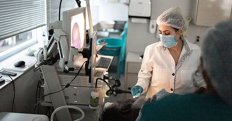
What is Creation of Pericardial Window or Partial Resection for Drainage: Overview, Benefits, and Expected Results
Definition & Overview
The process of creating a pericardial window is a method used by surgeons to drain excess fluid that has accumulated in the pericardium. The pericardium, also called the pericardial sac, is a double-layered sac that envelops the heart. It acts more like a cocoon that protects the heart from shock and friction from the surrounding organs.
Even though the pericardium is filled with fluid, especially the spaces within it (the pericardial cavity), excess fluid adds undue pressure to the heart leading to a condition called cardiac tamponade.
The heart’s performance is significantly impaired if it is subjected to too much pressure. When this happens, the heart will not be able to pump an adequate amount of blood to major arteries and veins. The entire process of blood oxygenation and blood transfer to arteries for distribution around the body will be significantly disrupted. This causes the entire human system to collapse since the whole body is dependent on oxygen for all its biological processes.
The creation of a pericardial window involves making an opening in the pericardial sac to drain out excess fluid trapped in and around the pericardial cavity. Surgeons surgically cut out some sections of the pericardium to allow the fluid to flow naturally into the chest cavity or around the peritoneum. The peritoneum is a membrane that makes up the lining of the abdominal cavity.
Other ways by which excess fluid can be drained out from the pericardium include the following:
- Subxiphoid incision – A minimally invasive method of inserting a catheter below the breastbone
- Thoracoscopy – A method that inserts an endoscope through a small incision in the chest
- Thoracotomy – An invasive surgical procedure that creates an incision in the chest wall
Who Should Undergo and Expected Results?
The creation of a pericardial window is indicated for:
- Symptomatic pericardial effusions
- Asymptomatic pericardial effusions, where the pericardial window is suggested to aid in making a definitive diagnosis
- Haemodynamically stable patients who do not show any outward circulation problems but are suspected of having pericardial effusion. In most cases, thoracoscopy is recommended.
- Patients exhibiting pericardial, pleural (anything that involves the lungs), and pulmonary diseases
- Recurrent effusions
- Tuberculous pericarditis, or the inflammation of the pericardium caused by the bacteria that causes tuberculosis. In this case, the creation of a pericardial window is important to prevent future tamponade or pericardial constriction.
- Removal of damaged tissue or foreign bodies from a wound especially in patients with mediastinitis, or the inflammation of the mediastinum (the tissues in the mid-chest)
- Effusions located in one or more fixed pockets in the pleural space. In medical science, this is conveniently called “loculated”.
- Chylopericardium pericardial effusion composed of chyle. Chyle is a bodily fluid with a milky appearance and is composed of fat droplets and lymph.
- Haemopericardium, or the presence of blood in the pericardial sac, which can also cause tamponade
- Patients with colonic or substernal gastric conduit. Patients with conduits are inserted with pipes or tubes where water, food, or human wastes can pass through temporarily.
Patients who undergo less invasive procedures usually require less recovery time. They can resume normal activities the day after while avoiding strenuous ones unless their surgeon advises otherwise.
For those whose chest walls were surgically opened, a longer hospital healing period is required. There’s no quick fix to shorten the recovery time for these patients.
Many doctors are still trying to find out the effectiveness of less invasive approaches in contrast to open surgeries. Many surgeons are starting to hypothesise that the best way to treat pericardial effusion is to create a permanent window in the pericardium through a more invasive thoracoscopy. They believe that the permanent window is the best solution to continuously drain fluid out from the pleural space and keep effusion from coming back.
How is the Procedure Performed?
Before the procedure starts, anaesthesic agent is applied. The patient will be put on IV and his or her vitals will be carefully monitored. A defibrillator is made available in case the patient’s heart stops beating during the procedure or develops arrhythmia or irregular heartbeat.
The removal of effusions in the pericardium using minimally invasive procedures only requires local anaesthesia. However, for thoracoscopic approaches or thoracotomy, general anaesthesia is required.
There are many methods used to create a pericardial window. These approaches vary depending on the accessibility of the pericardium and the overall haemodynamic stability of the patient. The patient can either be in a posterolateral position (just like when someone sleeps on their side) or supine, if the subxiphoid approach is to be used.
A short vertical incision is then made around 5-8 inches below the breastbone or the xiphoid process region. The xiphoid process is the lowest portion of the breastbone. A needle is inserted and eventually replaced by a catheter, which will be used to aspirate the fluid from the pericardial sac.
While the pericardium is being drained out, the innermost layer of the serous membrane of the pericardium is examined to look for any swelling or abnormal cells in the pericardial sac.
Thoracotomy and thoracoscopic approaches are invasive procedures and require the opening of the chest wall. Nevertheless, they often employ the same procedure in draining effusions from the pericardium.
Possible Risks and Complications
The procedure has high success and low recurrence rates compared to similarly popular procedures such as percutaneous pericardiocentesis or surgery.
However, some notable risks and complications may include bacterial infection, bleeding, irregular heartbeat or arrhythmia, myocardial infarction, cardiac arrest, or sudden death.
Such complications and risks rarely occur and should not be a hindrance for any patient to take advantage of the procedure. In the majority of cases, the benefits always outweighed the risks.
References:
Oxford Journals; “The pericardial window: is a video-assisted thoracoscopy approach better than a surgical approach?”; http://icvts.oxfordjournals.org/content/12/2/174.long
US National Library of Medicine National Institutes of Health; “Pericardial window procedures: Implications on left ventricular function”; http://www.ncbi.nlm.nih.gov/pmc/articles/PMC3605479/
/trp_language]
[trp_language language=”ar”][wp_show_posts id=””][/trp_language]
[trp_language language=”fr_FR”][wp_show_posts id=””][/trp_language]
## What is Creation of Pericardial Window or Partial Resection for Drainage?
### Overview
The creation of a pericardial window or partial resection is a surgical procedure that aims to drain excess fluid from the pericardial space, the area surrounding the heart. This fluid, known as pericardial effusion, can accumulate due to various medical conditions, leading to pressure on the heart and impaired cardiac function.
### Surgical Procedure
The surgery involves making an incision in the chest and creating an opening (window) in the pericardium, the fibrous sac enclosing the heart. Alternatively, a partial resection may be performed, where a portion of the pericardium is removed. The opening or resection allows the excess fluid to drain out, relieving pressure on the heart.
### Benefits
Creating a pericardial window or partial resection offers several benefits:
* Reduction of pericardial effusion and pressure on the heart
* Improvement of cardiac function and reduction of symptoms such as shortness of breath, chest pain, and fatigue
* Prevention of serious complications associated with untreated pericardial effusion, such as cardiac tamponade and heart failure
### Expected Results
The expected results of the surgery typically include:
* Improved cardiac function and reduced symptoms
* Resolution or improvement of pericardial effusion
* Prevention of complications and associated risks
### Key Considerations
Before undergoing the surgery, patients should discuss the following considerations with their healthcare provider:
* Potential risks and complications, such as bleeding, infection, and damage to surrounding structures
* Post-operative care and recovery time
* Long-term follow-up and monitoring
**Keywords:**
* Creation of pericardial window
* Partial pericardial resection
* Pericardial effusion
* Pericarditis
* Pericardial drainage
* Cardiac function
* Pericardial pressure


# PERICARDIAL WINDOW OR PARTIAL RESECTION FOR PERICARDIAL EFFUSION: OVERVIEW, BENEFITS, EXPECTED RESULTS