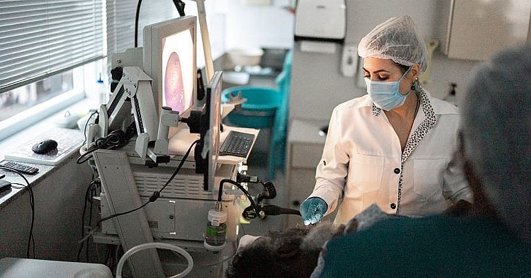
What is EEG: Overview, Benefits, and Expected Results
Definition and Overview
An electroencephalogram (EEG) is a type of test performed to measure the brain’s electrical activity to detect any irregularities indicative of different brain disorders. It is done by using special sensors, called electrodes, which are attached to the head and wired to a computer that records the brain’s electrical activity, which is represented as a series of wavy lines.
Who should undergo and expected results
An EEG is often performed to confirm the presence of the following conditions:
- Epilepsy
- Dementia
- Narcolepsy
- Problems in the nervous system
- Problems in the brain or spinal cord
- Mental health problems
Thus, patients showing signs of brain problems should undergo an EEG for a proper diagnosis. However, an EEG is also used for other purposes that are not diagnostic in nature. Aside from being a useful tool in the diagnosis of the disorders mentioned above, an EEG is also used to determine a patient’s chances of recovering from a comatose state as well as to monitor the brain activity of a person who is under anaesthesia.
The results of the electroencephalogram test are released on the same day the test is conducted or, at the latest, the day after. Depending on the pattern of the brain waves, a diagnosis can be made right away and the doctor will either declare the brain’s electrical activity as regular or abnormal. There are several types of brain waves that can show up on the test; these include:
- Alpha waves – This type has a frequency of 8 to 12 cycles per second. They occur only during the waking state or when one is mentally alert but has his or her eyes closed.
- Beta waves – With a frequency of 13 to 30 cycles, this type occurs when one is alert.
- Delta waves – This is the type that occurs when one is sleeping. This is also what can normally be found in young children.
- Theta waves – Similar to the Delta waves, this type occurs only when one is asleep. This has 4 to 7 cycles per second.
Normal brain activity means that the patient will have either alpha or beta waves while asleep and if both sides of the brain have similar brain wave patterns. The brain should also not have any sudden bursts of activity or an unexplained slowing down of electrical activity. At a certain point during the test, patients are asked to look at flashing lights to check the brain’s response; if the brain waves remain at a normal level, then the electrical activity is considered normal.
Brain activity is considered abnormal if the two sides of the brain have different wave patterns or if sudden spikes of electrical activity are detected. Likewise, if delta and theta waves are found when the patient is awake, then the brain’s activity is also considered as out of ordinary. Sudden spikes of activity are also something that doctors watch out for; these sudden spikes are associated with brain tumor, epilepsy, infection, or stroke. Its opposite is the absence of ongoing brain activity, which, in turn, indicates a comatose state.
Aside from determining if something is wrong, an EEG can also help determine the location of abnormal activity in the brain. This is vital in determining the type of epilepsy or seizure that the patient has. However, when using an EEG on a person suffering from epilepsy, it is important to remember that the results may appear normal in between seizure attacks.
How the procedure works
If you are scheduled to have an EEG, it is important to begin preparing for it a day before the test. The patient must avoid consuming anything that might have some effect on the brain’s electrical activity; these substances include sedatives, tranquilizers, sleep-inducing medication, coffee, tea, soda, and chocolates. The EEG test also requires patients to make sure that the scalp is clean, since the procedure requires several metal disks to be attached to it. So, avoid putting oil, conditioner, cream, or spray on the hair before going to the hospital for the test. Some doctors also suggest sleeping for a lesser number of hours before the test because the patient may be required to sleep while the test is being performed.
An EEG is usually done in a hospital under the supervision of an EEG technologist. With the patient lying down, the procedure begins with the technologist attaching several flat metal discs or electrodes to different spots on the patient’s head. These metal disks are attached to the head with the use of a sticky paste, or in some instances, needles. Sometimes, the individual small metal discs are replaced by one whole cap with electrodes already fixed on it. These electrodes are then attached to a computer where the brain’s electrical activity is recorded.
While the procedure is ongoing, the patient will be asked to lie still and will not be allowed to talk. The EEG technologist watches from a window and will communicate with the patient to ask him or her to do several tasks that will help in forming a diagnosis; these include:
- Breathing deeply and rapidly for 20 minutes
- Looking at a bright, flashing light
- Sleeping (if a patient finds it hard to fall asleep during the test, sedatives may be administered.)
The test usually lasts for 1 to 2 hours. However, in cases wherein the test is being performed to observe a sleep-related problem, the recording of brain activity may last for the whole duration of the patient’s sleep.
Possible risks and complications
An EEG test does not cause any pain to the patient. However, in cases where needles are used instead of paste, a prickling sensation may be felt while the needles are being inserted. If paste is used, some patients may have paste left on their hair for a while; this is the substance used to attach the electrodes to the scalp.
An electroencephalogram (EEG) is a very safe procedure with a very low risk of possible complications because no amount of electrical current will enter the body during the procedure. The only possible complications affect patients with seizure disorders, as the flashing lights that are part of the test may trigger a seizure attack. Thus, technologists who conduct the test use precautionary measures when performing it on a patient with epilepsy or other seizure-related disorders.
References:
- Salisbury D. “Clinical EEG and Neuroscience.” Journal of the EEG and Clinical Neuroscience Study.
- Song Y. (2011). “A review of developments of EEG-based automatic medical support systems for epilepsy diagnosis and seizure detection.” Scientific Research.
- Duffy F., Shankardass A. et al. (2013). “The relationship of Asperger’s syndrome to autism: a preliminary EEG coherence study.” BMC Medicine.
- British Medical Journal: “The EEG Apparatus.”
- Noor Kamal Al-Qazzaz, Sawal Hamid Bin Ali, Siti Anom Ahmad, et al. (2014). “Role of EEG as Biomarker in the Early Detection and Classification of Dementia.” The Scientific World Journal.
/trp_language]
[trp_language language=”ar”][wp_show_posts id=””][/trp_language]
[trp_language language=”fr_FR”][wp_show_posts id=””][/trp_language]
**Q: What is EEG?**
**A:** Electroencephalography (EEG) is a non-invasive procedure that records electrical activity in the brain using electrodes placed on the scalp. EEG patterns can vary depending on brain state, such as wakefulness, sleep, or altered consciousness.
**Q: What do EEG results show?**
**A:** EEG results typically display rhythmic patterns of brain activity, characterized by their frequency and amplitude. Different brain regions exhibit distinct frequencies, reflecting ongoing neural processes:
– **Delta waves** (0.5-4 Hz): Slow, high-amplitude waves associated with deep sleep.
– **Theta waves** (4-8 Hz): Associated with drowsiness, relaxation, and memory consolidation.
– **Alpha waves** (8-13 Hz): Present during wakefulness, relaxation, and meditation.
– **Beta waves** (13-30 Hz): Faster waves related to active thinking, problem-solving, and attention.
– **Gamma waves** (above 30 Hz): Brief, high-frequency bursts associated with sensory processing and complex cognitive functions.
**Q: How are EEG results interpreted?**
**A:** Interpretation of EEG results depends on factors such as age, medical history, and clinical context. Abnormal patterns or changes in EEG patterns can indicate underlying neurological conditions, including:
– Seizures and epilepsy
– Brain tumors
– Dementia and other neurodegenerative diseases
– Sleep disorders
– Brain trauma or injury
**Note:** It’s important to consult with a qualified healthcare professional, such as a neurologist or epileptologist, for the diagnosis and interpretation of EEG results.


## EEG: An Overview of Its Benefits and Expected Results