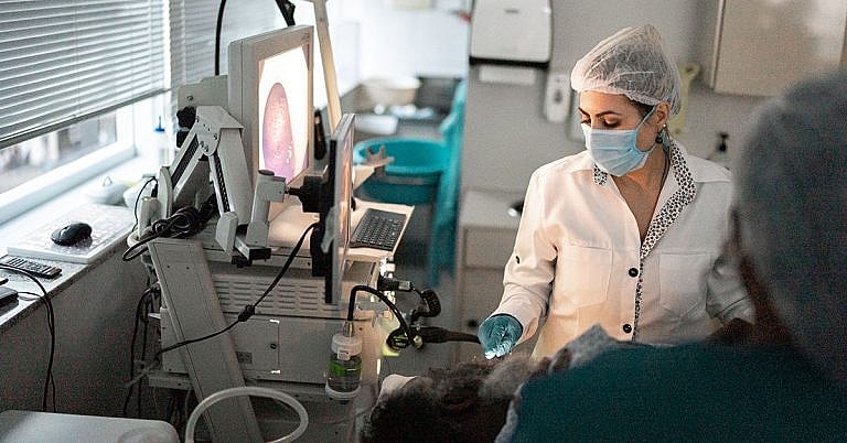
What is Endoscopic Transsphenoidal Surgery: Overview, Benefits, and Expected Results
Definition & Overview
An endoscopic transsphenoidal surgery is a minimally invasive procedure that removes pituitary tumours, particularly those in hard-to-reach areas of the brain, without the need for a large incision.
The procedure uses an endoscope, which is passed through the nostril to gain access to the transsphenoidal part of the brain, as well as special surgical instruments that are inserted through the endoscopic tube to remove the tumour.
The pituitary gland is a small structure located at the base of the brain and protected by the sphenoid bone, which is responsible for producing the hormones that help control the other glands of the body. Due to its role, the pituitary gland is also called the “master endocrine gland” in the body.
Pituitary tumours are usually small, benign, and slow-growing, but they can also become large and malignant. They can be removed through medication, radiotherapy, or surgery. Surgical options include conventional resection surgery and endoscopic transsphenoidal surgery.
Who Should Undergo and Expected Results
Endoscopic transsphenoidal surgery can be recommended to patients with abnormal growths on the pituitary gland, such as:
- Pituitary adenomas – These are non-cancerous tumours that grow slowly and do not spread to other body parts.
- Invasive adenomas – These are benign tumours that have the tendency to spread to the bones of the skull as well as to the sinus cavity just below the gland
- Pituitary carcinoma – These are malignant or cancerous tumours that can spread into other parts of the central nervous system, including the spinal cord and the brain.
- Craniopharyngioma – This is a benign tumour that grows near the pituitary stalk
- Rathke’s cleft cyst – This is a benign, fluid-filled growth that forms between the posterior and anterior lobes of the pituitary gland.
A transsphenoidal surgery may also be used for the removal of other brain tumours, such as meningioma and chordoma.
Pituitary tumours can be either functioning or non-functioning. Functioning tumours produce excessive amounts of hormones, which leads to the development of symptoms. Non-functioning tumours, on the other hand, are asymptomatic.
Symptoms that can accompany a functional tumour include:
- Headaches
- Vision problems
- Loss of body hair
- Loss of facial hair in men
- Abnormal breast growth in men
- Impotence in men
- Less frequent menstrual periods in women
- Inability to produce breast milk
- Reduced sexual libido
- Slowed growth in children
- Delayed sexual development
The risk of getting pituitary tumours is higher among those who have certain genetic conditions, such as multiple endocrine neoplasia type 1 syndrome, isolated familial acromegaly, or Carney complex, among others. The tumours are diagnosed using imaging, blood, and urine tests.
By getting rid of the tumour, an endoscopic transsphenoidal surgery is expected to fully treat the patient’s medical problem and relieve any related symptoms, as long as the tumour is benign. The resected tumour is typically placed under laboratory analysis following the procedure. If the tumour is found to be malignant, the patient is more likely to be advised to seek further cancer treatment.
The development of endoscopic transsphenoidal surgery has advanced the treatment of pituitary tumours, as the minimally invasive technique provides surgeons with direct access to the tumours without the need for a massive high-risk brain surgery. The procedure is performed without any incisions in the scalp and with minimal blood loss. It is effective in removing even tumours as large as 5 cm in diameter without affecting the pituitary gland.
How is the Procedure Performed?
The term “transsphenoidal” means “through the sphenoid sinus” indicating that the procedure is performed through the nose and sinus. It is also sometimes termed as an endonasal procedure. During surgery, an endoscope, which is a thin hollow tube, is passed through either the right or the left nostril, depending on where the tumour is located.
Such a procedure is performed by a neurosurgeon with the assistance of an ear, nose, and throat surgeon. Prior to the procedure, the patient will meet with the surgeon to discuss the surgery, its risks and benefits, and the patient’s medical history. It is important for the patient to provide a complete list of his allergies, medications, vitamins, reactions to anaesthesia, and previous surgical procedures so that the surgeon can take these into consideration in planning the procedure. The patient will also undergo pre-surgical tests, such as blood tests, ECG, chest x-ray, and CT scan.
On the day of the surgery, the patient is placed under general anaesthesia and an antibiotic and antiseptic solution is applied to the nasal cavity. An image guidance system is also placed on the patient’s head to be used by the surgeon while navigating through the nasal passages.
To start the procedure, the ENT surgeon proceeds to insert an endoscope into one of the nostrils and gradually advance it towards the back of the nasal cavity. The surgeon then passes long surgical instruments through the nostril. If necessary, a part of the nasal septum is removed. Once the sphenoid sinus is reached, the surgeon opens its front wall to reveal the sella, or the bone overlying the pituitary gland, to gain direct access to the lining of the skull or the dura. When the dura is opened, the surgeon will gain access to the pituitary gland and the tumour on it.
At this point, the neurosurgeon takes over to remove the tumour using special surgical instruments. First, the centre of the tumour is removed. Doing so causes the margins of the tumour to fall inwards, making it easier to reach and remove through the endoscope. Once the tumour is removed, the surgeon inspects the gland for more signs of disease or abnormal growths. When none is found, he proceeds to close the sella, sometimes using a small piece of fat graft taken from the abdomen and a bone graft taken from the septum to do so.
After the procedure, the patient is taken to a recovery room, where he will be placed under close monitoring until the anaesthesia wears off. An MRI scan will be performed the next day. The patient may need to stay in the hospital for 1 to 2 days after the procedure.
Possible Risks and Complications
Any surgical procedure has risks, especially one that involves a part of the brain. Due to the location of the pituitary gland, the procedure may cause:
- Vision loss
- Diabetes insipidus
- Meningitis
- Sinus congestion
- Nasal bleeding
- Nasal deformity
- Stroke
- Cerebrospinal fluid leakage
Damage to the pituitary gland
ReferencesWen G., Tang C., et al. “Mononostril versus binostril endoscopic transsphenoidal approach for pituitary adenomas: A systematic review and meta-analysis.” April 28, 2016. http://journals.plos.org/plosone/article?id=10.1371/journal.pone.0153397
Gao Y., Zhong C., Wang Y. “Endoscopic versus microscopic transsphenoidal pituitary adenoma surgery: a meta-analysis.” World J Surg Oncol. 2014; 12:94. http://www.ncbi.nlm.nih.gov/pmc/articles/PMC3991865/
/trp_language]
[trp_language language=”ar”][wp_show_posts id=””][/trp_language]
[trp_language language=”fr_FR”][wp_show_posts id=””][/trp_language]
**Q: What is Endoscopic Transphenoidal Surgery (ETS)?**
**A:** ETS is a surgical procedure used to access and operate on the pituitary gland, which is located at the base of the brain. It involves inserting an endoscope, a thin, flexible instrument with a camera attached, through the nose and into the sphenoid sinus, a bone located behind the nose. This technique allows for a less invasive approach compared to traditional surgery.
**Q: What are the Benefits of ETS?**
**A:**
– **Non-invasive:** ETS does not require any external incisions or cutting, leading to reduced pain and scarring.
– **Shorter recovery time:** Patients typically recover from ETS within a few days, with minimal post-operative pain.
– **Preservation of normal structures:** The endoscopic approach allows surgeons to visualize and avoid damage to surrounding tissues.
- **Cosmetically appealing:** Since it doesn’t involve any external incisions, ETS leaves no visible scars.
**Q: What are the Expected Results of ETS?**
**A:** The outcomes of ETS vary depending on the underlying condition being treated. However, the expected results generally include:
– **Improved pituitary function:** ETS can effectively remove tumors or cysts from the pituitary gland, restoring hormonal balance.
– **Reduced symptoms:** For patients with conditions such as Cushing’s disease or prolactinomas, ETS can alleviate symptoms and improve quality of life.
– **Preservation of vision:** ETS can help prevent further damage to the optic nerves, which can occur in conditions like pituitary tumors.
– **Long-term remission:** In many cases, ETS can lead to long-term remission or significant improvement in the underlying condition.
**Q: Is ETS the Right Surgery for you?**
**A:** The decision of whether ETS is the right surgery for you depends on several factors, including your specific condition, your medical history, and your preferences. It’s essential to have a thorough consultation with a qualified ear, nose, and throat (ENT) specialist or neurosurgeon to determine the best approach for your case.


This is a great article. I learned a lot about endoscopic transsphenoidal surgery. Thank you for sharing!
Great article