What is Fluoroscopy: Overview, Benefits, and Expected Results
Definition & Overview
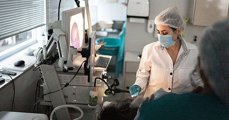
A fluoroscopy is a medical imaging procedure that allows doctors to capture still images of specific organs and to observe a video showing the movements of different body parts on a fluorescent screen in real-time. This procedure uses X-ray technology and contrast dye material, which makes the targeted body parts radio-opaque for easier visualization. It is commonly used in the diagnosis of diseases and also as an interventional procedure in the fields of orthopaedic, gastroenterology, and cardiovascular care.
Who should undergo and expected results
Fluoroscopy is typically used for diagnosing medical conditions in the fields of:
- Gastroenterology – Fluoroscopy procedures for gastrointestinal monitoring commonly use barium as a contrast agent to observe the movements and assess the condition of gastrointestinal organs, which include the esophagus, stomach, large intestine, and small intestines to find the cause of gastrointestinal symptoms, such as vomiting, trouble swallowing, belly pain, or indigestion. It can also be used to find polyps, tumours, or to confirm the presence of malabsorption syndrome.
- Orthopedic – Fluoroscopy is most commonly used in the field of orthopedic to monitor the healing of broken bones to ensure that they are properly positioned and aligned throughout the healing process. It is also performed to assist in the insertion of implants.
- Cardiovascular Care – Cardiovascular fluoroscopies are usually used when a blockage is suspected; the procedure can assist in the insertion and progression of a catheter used to diagnose and treat the condition.
A fluoroscopy is expected to provide doctors with information that is otherwise impossible to obtain using other tests. This information is used to determine the right course of action in terms of treatment or to determine whether further action is necessary, in cases of monitoring procedures.
How the procedure works
Fluoroscopy procedures are routine imaging tests that usually take 45 minutes to an hour, although the length of each procedure may vary depending on the part of the body that is being assessed. The process usually begins with the application of a contrast dye material. If used for gastrointestinal imaging, this part of the procedure may cause some discomfort as the patient has to swallow the said material. As it travels through the GI tract, the doctors obtain clear images of the esophagus, stomach, small intestines, and large intestines. A contrast dye may also be used for the examination of the rectum, but it is placed into the body using an enema tube instead of making the patient swallow it.
The preparations needed for the procedure are simple. Once a patient arrives at the designated imaging area, he or she will first be asked to change to a lab gown. The procedure proceeds with the injection of the anesthesia or sedation. There are some instructions regarding the use of anesthesia and these are typically sent to the patient days before the actual procedure. This list of instructions must always be reviewed and followed carefully.
Once preparations are in place, the fluoroscopy scan begins. There are two types of equipment that can be used in this procedure, fixed systems and a mobile alternative. Fixed systems are used in designated imaging labs, while the C-arm mobile fluoroscopic unit provides flexibility in terms of where the procedure can be conducted.
The actual procedure uses x-ray beams, which create a representation of the body’s interiors as it passes through at a maximum rate of 25 to 30 frames per second, making video representations possible. The output passes through special equipment that helps intensify and brighten the images before they are finally transmitted to a fluorescent screen. More recent models of the equipment make it possible to digitize the pictures.
Possible risks and complications
Like most medical procedures, fluoroscopy has associated risks that are primarily caused by radiation. This is the reason why it is not prescribed for pregnant women due to the potentially harmful effects of radiation on a developing fetus. As a rule, the imaging test is only used if its intended benefits outweigh the potential risks.
As much as possible, medical professionals use low dose of radiation to minimize the risks. However, this depends on the condition of the patient. In cases where fluoroscopy is used to aid lengthy procedures (such as in interventional procedures that require the insertion of stents), the dose is adjusted accordingly, so there is a possibility that the radiation that a patient will receive will be relatively high.
Risks involved with a fluoroscopy falls under two types, namely deterministic and stochastic. These risks include:
- Injuries to the skin and tissues such as burns
- Cataracts due to the radiation
- Cancer
Aside from the radiation, there are also other elements in the procedure that can cause undesired effects such as complications that may arise from the use of anesthesia or sedation.
To minimize the risks associated with the procedure, medical professionals must check the:
- Cumulative amount of radiation exposure that the patient is subjected to
- Size of the x-ray field, which can be reduced so that the x-ray will only be applied within the image target area and will not affect nearby parts of the body
- Exact positioning of the x-ray to eliminate the need for repeat exposures
- X-ray filtration, which is especially important in long procedures
Special last-image hold feature for viewing an image repeatedly without continuous patient exposure
References:Rockville, MD. Food and Drug Administration. Public Health Advisory: Avoidance of Serious X-Ray-Induced Skin Injuries to Patients During Fluoroscopically-Guided Procedures. Center for Devices and Radiological Health, FDA. 1994.
Wagner LK, Eifel PJ, Geise RA. Potential biological effects following high X-ray dose interventional procedures. J Vasc Interv Radiol. 1994 Jan-Feb. 5(1):71-84.
Huda W, Peters KR. Radiation-induced temporary epilation after a neuroradiologically guided embolization procedure. Radiology. 1994 Dec. 193(3):642-4.
/trp_language]
[trp_language language=”ar”][wp_show_posts id=””][/trp_language]
[trp_language language=”fr_FR”][wp_show_posts id=””][/trp_language]
**What is Fluoroscopy?**
**Overview**
Fluoroscopy is a medical imaging technology that generates real-time moving X-ray images. It allows doctors to visualize internal structures and their movement inside the body in real-time. Unlike conventional X-rays, fluoroscopy provides a continuous stream of images rather than a single static snapshot.
**Benefits of Fluoroscopy**
* **Real-Time Visualization:** Fluoroscopy eliminates the need for multiple X-ray exposures, offering continuous imaging for precise diagnosis and treatment.
* **Dynamic Examination:** Dynamic fluoroscopy enables the visualization of moving structures, such as the heart beating or the joints in motion, providing valuable diagnostic information.
* **Safety:** Fluoroscopy delivers lower radiation dosage compared to traditional X-ray techniques, minimizing patient exposure.
* **Intraoperative Visualization:** Fluoroscopy assists during surgical procedures, providing surgeons with real-time images to guide and navigate their interventions.
* **Diagnostic and Therapeutic Procedures:** Fluoroscopy facilitates both diagnostic procedures (e.g., cardiac catheterization) and therapeutic interventions (e.g., balloon angioplasty).
**Expected Results**
Depending on the examination, fluoroscopy can provide detailed images of various body parts and organs, including:
* **Bones:** To evaluate fractures, joint disorders, or tumor growth.
* **Gastrointestinal Tract:** To assess the function of the esophagus, stomach, and intestines (barium swallow, upper GI series).
* **Cardiovascular System:** To diagnose heart conditions, check blood flow, and assess vascular structures (cardiac catheterization, angiography).
* **Lungs:** To detect lung abnormalities or fluid accumulations (fluoroscopic CT).
* **Urinary Tract:** To diagnose urinary tract conditions or pain (intravenous pyelogram, voiding cystourethrogram).
* **Reproductive System:** To evaluate infertility issues or uterine problems (hysterosalpingogram).
**Conclusion**
Fluoroscopy offers a wide range of diagnostic and therapeutic applications. Its real-time capabilities and lower radiation dosage make it an invaluable tool for healthcare professionals. By providing detailed and dynamic images of internal structures, fluoroscopy enhances patient care, enables precise diagnosis, and assists in effective medical interventions.


One comment