
What is Head CT Scan: Overview, Benefits, and Expected Results
Definition & Overview A CT scan of the head, also called a cranial scan, is an advanced x-ray technology that takes images of the head. A CT scan or computed...
Definition & Overview
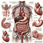
A CT scan of the head, also called a cranial scan, is an advanced x-ray technology that takes images of the head. A CT scan or computed tomography scan, however, is not limited to head scans; it can be used to take images of any part of the body, providing doctors with clear pictures of a patient’s internal organs and bones. Its main advantage over traditional x-ray is its ability to provide detailed images of the blood vessels and soft tissue found in the head. This makes it a more accurate scanning technology that now plays a major role in the diagnosis of countless head and brain conditions.
Who Should Undergo & Expected Results
A CT scan of the head delivers information that is vital to the evaluation of the brain. It can also provide crucial information regarding the different parts of the face, such as the eyes, nasal structure including the sinuses, facial bones, and even the inner ear. However, the information provided is very limited, as cranial scans are focused more on the brain. If specific parts of the face are in question, other more specific types of scans may be conducted.
Due to the type and scope of information it delivers, a CT head scan is most commonly performed on people who are suspected of having brain injury, brain tumour, internal bleeding in the brain, or aneurysm. The CT scan is recommended for patients who are experiencing the following symptoms:
- Headaches
- Confusion
- Numbness
- Vision problems
- Vertigo
It is also necessary for patients who have suffered from paralysis or stroke, as a CT scan also shows the extent of damage that these conditions have caused.
The information received from a CT scan of the head helps physicians to:
- determine the best and most appropriate treatment for the patient
- plan for surgery
- evaluate the results of previously conducted treatment or surgery
In a CT scan of the head, the following guide is used to evaluate the results.
In a normal CT scan, the brain, blood vessels, and the skull bones should all have normal size and positioning, with no foreign growths and no inflammation, bleeding, or clots. On the other hand, results are considered abnormal if there is (1) an abnormal growth, tumour, or bleeding in or around the brain (2) presence of any foreign objects such as glass or metal (3) any broken bones in the skull or pinched nerves leading to and from the brain (4) collected fluid, aneurysm, swelling or edema, and (5) enlarged brain ventricles.
How Does the Procedure Work?
A radiology technologist performs the CT scan of the head, while its results are written and analyzed by a radiologist. Other medical professionals, however, are also equipped to review and analyze the printed results of a CT cranial scan.
In preparation for the scan, patients are asked to remove any jewelry, glasses, or hearing aids they may have on their person. They are also advised to wear only loose-fitting clothes that are comfortable to wear.
During the scan, the patient will be asked to lie on a table attached to the CT scanner, which is then positioned over the head. Straps may be placed around the head to prevent any unnecessary movements. Before the scan begins, the scanner will let out a click or buzz. The scanner then takes x-ray images of the head, with the scanner rotating and tilting to scan from different angles and positions. The images are then sent to a computer for analysis and printing. To obtain accurate and clear pictures, patients are asked to lie very still during the scan, which lasts for only a few seconds. However, the entire CT test may take up to 60 minutes in total, with most of the time spent preparing you for the scan. Radiologists then take up to 1 to 2 days to release the results to the requesting physician.
When called for, radiologists also use a dye-based contrast material that is made to flow through the veins. The contrast material makes it easier to identify the different parts of the head on a CT image, and is very effective in checking for blood flow around the brain, abnormal growths and tumours, inflamed areas, or nerve damage.
Take note, also, that patients are left alone in the scan room during the test, and the technologist or radiologist performing the scan will only watch from a window. The scan room is equipped with a two-way intercom to allow the patient and technologist to communicate.
CT scans of the head can also be performed on children, but they will be given special instructions and will need their patients’ or guardians’ support and cooperation during the scan. Children that are too young may feel afraid or may not be able to hold still for the scan may be given sedatives with the consent of their parents.
Possible Complications and Risks
Although a computed tomography scan of the head will not cause any pain, it does have some possible complications and risks. As a radiation-based diagnostic scan, there is a very low risk of cancer, especially in patients who undergo a lot of radiation tests. If this is a concern, especially among paediatric patients, the attending physician should be consulted regarding the risk of radiation exposure and the need to conduct the scan.
And since, like traditional x-ray scans, a CT scan uses x-ray technology, the same limitations and precautions apply. Thus, patients should inform their attending physician if they:
- are pregnant or might be pregnant
- have allergies to medications and iodine-based dyes
- have a heart condition
- are suffering from diabetes or are taking medications for diabetes such as metformin (This is usually resolved by adjusting the schedule of your medication. Patients are usually first given a schedule when to stop taking the drugs prior to the scan and when to restart medication after the test.)
- have kidney problems
- are suffering from asthma
Some patients may feel nervous or uncomfortable due to the confined space inside the CT scanner. In such cases, sedatives may be administered to help them relax.
References:
- Shaw AS, Dixon AK. Multidetector computed tomography. In: Adam A, Dixon AK, eds. Grainger & Allison’s Diagnostic Radiology: A Textbook of Medical Imaging. 5th ed. New York, NY: Churchill Livingstone; 2008:chap 4.
/trp_language]
**What is a Head CT Scan?**
A Head Computed Tomography (CT) scan, also known as a brain scan or head scan, is a non-invasive imaging test that uses X-rays and computer processing to create detailed cross-sectional images of the head. It provides valuable information about the brain, skull, and surrounding structures.
**Overview of Head CT Scan**
* Uses X-ray technology and computerized image processing
* Produces cross-sectional images or “slices” of the head
* Provides detailed views of brain tissue, skull, and other anatomical structures
* Can be performed with or without contrast enhancement (involving injection of a contrasting agent)
**Benefits of a Head CT Scan**
* Fast and widely available imaging modality
* Detects and diagnoses a wide range of neurologic conditions
* Emergency evaluation of head injuries, strokes, and bleeding
* Assessment of brain structures, anatomy, and abnormalities
* Identification of tumors, cysts, and infections
* Pre-operative planning and surgical guidance
**Expected Results of a Head CT Scan**
The results of a head CT scan are typically interpreted by a radiologist, who will generate a report that describes the findings. Expected results may include:
* Normal brain anatomy and structures
* Presence of any abnormalities, such as:
* Tumors
* Hemorrhages
* Strokes
* Fractures
* Infections
* Evaluation of intracranial bleeding, swelling, or herniation
* Assessment of blood flow and perfusion through the brain
**Tips for Preparing for a Head CT Scan**
* Inform your healthcare provider about any medical conditions or medications
* Remove all jewelry and metallic objects before the scan
* Be prepared for administration of a contrast agent if necessary
* Follow specific instructions provided by the imaging center
**Conclusion**
A head CT scan is a valuable imaging tool that provides crucial information about the head and brain. It is often used as a first-line diagnostic test for a variety of neurologic conditions. By understanding the overview, benefits, and expected results of a head CT scan, patients can prepare themselves for the procedure and fully utilize the information it provides.
One comment
Leave a Reply


Popular Articles


What is a Head CT Scan, Overview, Benefits, and Expected Results
This title accurately reflects the intent and content of the post.