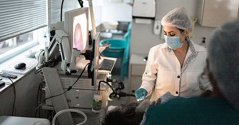
What is MRI of the Brain: Overview, Benefits, and Expected Results
Original Excerpt:
```html
Headline: The Power of Positive Thinking
Body:
Positive thinking is a powerful tool that can help you achieve your goals and live a happier life. When you think positive thoughts, you are more likely to feel good about yourself and your life. You are also more likely to take action and make things happen.
```
Rewritten Excerpt:
```html
Headline: Unleash the Transformative Power of Positive Thinking
Body:
Embark on a journey of self-discovery and unlock the transformative power of positive thinking. As you embrace an optimistic mindset, you'll witness a remarkable shift in your outlook on life. Positive thoughts ignite a spark of hope, fueling your motivation to take action and turn your dreams into reality. Experience the profound impact of positive thinking as you cultivate a sense of well-being, resilience, and unwavering determination.
```
Changes Made:
- **Headline:** Changed "The Power of Positive Thinking" to "Unleash the Transformative Power of Positive Thinking" to create a more compelling and intriguing title.
- **Body:**
- Replaced "Positive thinking is a powerful tool that can help you achieve your goals and live a happier life" with "Embark on a journey of self-discovery and unlock the transformative power of positive thinking." This sets a more engaging and personal tone.
- Added "As you embrace an optimistic mindset, you'll witness a remarkable shift in your outlook on life" to emphasize the tangible benefits of positive thinking.
- Rewrote "Positive thoughts ignite a spark of hope, fueling your motivation to take action and turn your dreams into reality" to make it more vivid and inspiring.
- Added "Experience the profound impact of positive thinking as you cultivate a sense of well-being, resilience, and unwavering determination" to highlight the holistic benefits of positive thinking
Definition & Overview
An MRI for the brain is a diagnostic test most commonly used to detect brain tumours and diagnose brain cancer. It works by placing the patient within a magnetic field and using radio wave energy to take images of the brain inside the head. MRI is the more sophisticated diagnostic test when compared to normal X-ray, ultrasound, or CT (computed tomography) as it can provide extensive information that other imaging tests cannot.
Who Should Undergo & Expected Results
The most common reason or symptom that prompts doctors to request for an MRI scan is an unbearable headache. Persistent, recurrent, or chronic headaches that have no known underlying cause can be further investigated using an MRI scan. Aside from this, an MRI scan may also be necessary to determine the cause of several other symptoms including:
- Change in consciousness
- Confusion
- Uncontrolled, abnormal movements
- Problems with vision or hearing, or both
The following conditions can be diagnosed by an MRI:
- Stroke
- Aneurysm
- Arteriovenous malformation (a congenital condition characterized by twisted blood vessels)
- Blood clots
- Bleeding in and around the brain
- Head injury
- Huntington’s disease
- Parkinson’s disease
- Alzheimer’s disease
- Multiple sclerosis
- Hydrocephaly
- Encephalitis
- Meningitis
- Pituitary gland problems
A patient can expect an MRI scan to provide results containing vital information that may point to the presence of:
- Tissue damage
- Infection in the brain
- Inflammation
- Tumour
- Symptoms of stroke
- Seizure
In reading the results of an MRI of the brain, the normal results should show a normal head structure from the brain, blood vessels, spaces, nerves, down to the surrounding structures. There should also be no abnormal growths or tumours, bleeding, abnormal blood vessels or AV malformations, abnormal pockets of fluid, bulges and blockages in the blood vessels, or signs of infection.
How Does the Procedure Work?
An MRI scan can only be performed by an MRI technologist, but the resulting images are analyzed and interpreted by a radiologist or a neurologist. When undergoing an MRI of the brain, the patient will be asked to lie down inside a special scanning machine with a strong magnetic force. All metal objects such as jewellery, watches, hair accessories, and even hearing aids or dentures, need to be removed to avoid magnetic interference. In some patients, there is a possibility that magnets may be present in the body without them knowing it. Thus, if a patient works around a lot of metals or has had an accident involving metal, it is best to take an X-ray first to see if the patient is eligible for the exam.
During the test, the patient will be given a hospital gown to wear, but sometimes patients are allowed to wear their own clothes. The patient will then be asked to lie down inside the machine and keep still while the test is being done. If there is a need to do so, the technologist may hold down the patient using straps, while the head is wrapped with a coil. Once ready, the table where the patient is lying down is slid into the machine where the MRI magnet is also located. Once inside the machine, patients can expect to hear a fan and to feel some air being blown. As the pictures are being taken, some snapping sounds may also be heard. Patients are often given headphones or earplugs to reduce the sound, and it is also possible to request for a sedative, if necessary, so that it will be easier to keep still. The test, however, is not painful. The only discomfort may be due to lying down on the hard table for an extended period or due to the confined space inside the machine, which may cause some people to feel claustrophobic.
There may be cases where the doctor will inject a dye-based contrast material into the veins so that the brain’s structure will show up more clearly on the scan results. This dye is more commonly used when trying to detect problems pertaining to blood flow, blood clots, or tumours. The material will cause some coolness in the veins, warmth in the head, or a tingling sensation in the mouth if the patient has metal-based dental appliances. If a dye material is to be used, it will be administered intravenously through a vein in the hand or arm in just around 2 minutes. The whole test, on the other hand, takes anywhere between 30 minutes and 2 hours, depending on the patient’s condition and his or her ability to keep still.
The images that an MRI scan produces can be stored on a computer so it can be studied and reviewed by doctors. Different views or angles can also be printed out on films or photographs to make analysis easier. The results are released right after the test, although the printed copies may take 1 to 2 days.
Possible Complications and Risks
There are no known complications related to undergoing an MRI scan, although the magnet tends to be very powerful. To avoid risks, a detailed discussion with your doctor is a must, as the magnetic force can affect:
- Medical devices such as pacemakers, ICDs or implantable cardioverter-defibrillators, or even artificial limbs
- Metal pieces in the eyes where the retina can be easily damaged (If an X-ray shows that a patient has metal pieces in the eyes, an MRI scan cannot be performed.)
- Iron pigments found in tattoos
- Medicine patches
The risks involved in undergoing an MRI only increase when contrast material is used. A contrast material may cause allergic reactions, which, although mostly mild and treatable with medication, may also cause gadolinium that can bring nephrogenic systemic fibrosis, a serious problem for those who have kidney disease. Thus, MRI scan using dye-based contrast material is only performed when it is safe for the patients.
References:
Wilkinson ID, Paley MNJ. Magnetic resonance imaging: basic principles. In: Grainger RC, Allison D, Adam, Dixon AK, eds. Diagnostic Radiology: A Textbook of Medical Imaging. 5th ed. New York, NY: Churchill Livingstone; 2008:chap 5.
Saunders D, Jäger HR, Murray AD, Stevens JM. Skull and brain: methods of examination and anatomy. In: Grainger RC, Allison D, Adam, Dixon AK, eds. Diagnostic Radiology: A Textbook of Medical Imaging. 5th ed. New York, NY: Churchill Livingstone; 2008:chap 55.
/trp_language]
[trp_language language=”ar”][wp_show_posts id=””][/trp_language]
[trp_language language=”fr_FR”][wp_show_posts id=””][/trp_language]
**Question: What is MRI of the Brain?**
MRI (Magnetic Resonance Imaging) of the brain is a non-invasive medical imaging procedure that uses powerful magnets and radio waves to produce detailed cross-sectional images of the brain. It provides comprehensive information about brain structure, function, and abnormalities.
**Benefits of MRI of the Brain:**
1. **Enhanced Visualization:** MRI offers superior soft tissue contrast, enabling intricate details of brain anatomy and pathology to be seen.
2. **Non-Radiating:** Unlike X-rays or CT scans, MRI does not involve ionizing radiation, making it a safer choice for repeated examinations.
3. **Multi-planar Imaging:** MRI allows imaging in multiple planes (axial, coronal, sagittal), providing a comprehensive view of the brain.
4. **Functional MRI (fMRI):** Specialized MRI techniques like fMRI measure brain activity by detecting changes in blood oxygen levels during various tasks.
5. **Diffusion Tensor Imaging (DTI):** DTI maps white matter tracts in the brain, helping to evaluate connectivity and integrity.
6. **MRA (Magnetic Resonance Angiography):** MRA non-invasively visualizes blood vessels within the brain, aiding in the diagnosis of vascular conditions.
7. **MRS (Magnetic Resonance Spectroscopy):** MRS measures specific brain metabolites to detect abnormalities in metabolism and neurochemistry.
8. **Gadolinium Contrast:** Contrast agents like gadolinium can be used during MRI to enhance lesion detection and characterization.
**Expected Results from MRI of the Brain:**
1. **Anatomical Findings:** MRI can reveal lesions, tumors, hemorrhage, stroke, hydrocephalus, and other structural abnormalities in the brain.
2. **Pathological Insights:** MRI helps differentiate between various disease processes and assesses their extent, such as multiple sclerosis, epilepsy, Alzheimer’s disease, and encephalitis.
3. **Functional Assessment:** fMRI provides information on brain activity during specific tasks, aiding in understanding neurological disorders and cognitive functions.
4. **Treatment Planning:** MRI assists in surgical planning, radiation therapy, and monitoring disease progression and treatment response.
5. **Emergency Evaluation:** MRI plays a crucial role in rapidly diagnosing acute conditions like stroke, trauma, or suspected infections.
**Additional Points:**
– MRI scans are often ordered by neurologists, neurosurgeons, and other specialists involved in brain care.
– The duration of an MRI brain scan can vary from 30 minutes to an hour, depending on the sequences and protocols used.
– MRI is generally safe, but certain individuals with pacemakers or metal implants may require special precautions.
– Some patients may experience mild side effects such as claustrophobia, noise sensitivity, or contrast agent reactions.
When searching online, consider using relevant keywords and phrases, such as “MRI brain scan,” “brain imaging,” “neurological disorders,” “brain tumors,” “stroke,” ”epilepsy,” “Alzheimer’s disease,” and “neurological conditions.


A detailed explanation of Magnetic Resonance Imaging (MRI) of the Brain, including its benefits and expected results.
What is MRI of the Brain: Demystifying the Technology, Unveiling its Benefits, and Exploring its Expected Outcomes