What is Resection of Lateral Pharyngeal Wall: Overview, Benefits, and Expected Results
The new product is a great addition to our lineup.
Our latest product is an exciting addition to our already impressive lineup! With its innovative features and sleek design, it's sure to be a hit with customers. Don't miss out on this amazing opportunity to upgrade your life!
Overview of Resection of Lateral Pharyngeal Wall
Resection of the lateral pharyngeal wall is a minimally invasive surgery used to treat sleep apnea and breathing problems associated with certain neurological conditions. This type of surgery can help improve airway resistance, reduce snoring, and restore the natural airway anatomy.
The procedure involves the resection (removal) of the lateral part of the pharyngeal wall (the throat), which is located in the neck area. The goal of the surgery is to remove tissue blocking the airway of the patient and allow air to flow through without obstruction.
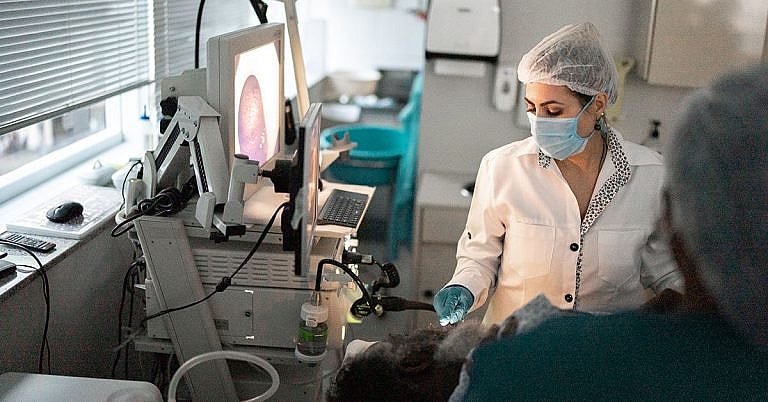
Surgeons often perform the procedure using an endoscope, which is an instrument that has a camera and light on the tip. The surgeon inserts the endoscope into the throat of the patient under general anesthesia and then begins to remove tissue from the lateral pharyngeal wall. Depending on the type and location of the obstruction, the surgeon may remove just a small portion of tissue or up to a few centimeters of tissue.
The procedure usually takes about one hour to complete and is typically an outpatient procedure. Additionally, the patient may be expected to stay in the hospital for one or two days for observation and will be able to return home once they are fully recovered.
Benefits of Resection of Lateral Pharyngeal Wall
The primary benefit of the procedure is that it can help improve airway resistance, reduce snoring, and restore the natural airway anatomy. In addition to improved breathing, resection of the lateral pharyngeal wall can also improve sleep quality and reduce daytime fatigue. Additionally, it can reduce the risk of sleep apnea related complications, such as high blood pressure and stroke.
Patients who undergo this procedure often have an improved quality of life and may be able to sleep better, have more energy throughout the day, and have an improved mood. In addition, the surgery is minimally invasive, so it generally has a short recovery time and minimal discomfort.
Expected Results After Resection of Lateral Pharyngeal Wall
The expected results after resection of the lateral pharyngeal wall are an improved quality of life and improved airway anatomy. Depending on the extent of the obstruction, some patients may be able to eliminate their obstructive sleep apnea, while others may reduce the obstructive sleep apnea significantly.
After the surgery, the patient may be expected to have some soreness or discomfort in the throat area, but this should subside with time. Additionally, the patient may experience some swelling, but this too should subside over time.
In most cases, patients will experience an improvement in breathing within the first few weeks after the procedure, but the full results may not be apparent for several months. The patient should follow up with their doctor to ensure the surgery was successful and that the airway is clear.
Potential Complications of Resection of Lateral Pharyngeal Wall
While the resection of the lateral pharyngeal wall is generally a safe procedure, there are some potential complications associated with it. These include infection, bleeding, scarring, and damage to the surrounding tissue. Additionally, the procedure may not provide adequate relief in some cases, so the patient may require additional procedures or therapies to completely resolve their breathing issues.
Patients should speak with their doctor to discuss the potential risks and benefits of this procedure in order to make an informed decision.
Summary
Resection of the lateral pharyngeal wall is a minimally invasive surgery used to treat sleep apnea and breathing problems associated with certain neurological conditions. The goal of the surgery is to remove tissue blocking the airway of the patient and allow air to flow through without obstruction. The primary benefit of the procedure is that it can help improve airway resistance, reduce snoring, and restore the natural airway anatomy. The expected results after the procedure are an improved quality of life and improved airway anatomy. While the resection of the lateral pharyngeal wall is generally a safe procedure, there are potential risks and benefits that should be discussed with a doctor before undertaking the procedure.
Definition & Overview
The resection of the lateral pharyngeal wall is a surgical procedure of removing tumours or cancerous growths from the lateral wall of the pharynx. Also known as pharyngotomy, the procedure is often performed to treat carcinoma of the oropharynx and pyriform sinus.
The oropharynx is located right behind the oral cavity or the mouth. It starts from the uvula and ends in the hyoid bone. The lateral wall of the oropharynx is where the palatine tonsils are located. Other parts of the lateral wall are the tonsillar fossa and tonsillar pillars. The whole oropharynx is part of the pharynx that functions as a passageway for food and air.
The pyriform sinuses are a pair of recessed structure found on the sides of the larynx. It is also called the pyriform recess, the pyriform fossa, smuggler’s fossa, or vallecula of the throat. It is also where the internal laryngeal nerve is located.
Who Should Undergo and Expected Results
Patients diagnosed with oropharyngeal cancer, especially those in the lateral pharyngeal wall, are eligible for surgery. This condition typically affects mature adults and occurs more often in men than women. Among its risk factors are a long history of smoking and an infection caused by a particular strain of the human papillomavirus. This condition is characterised by pain in the neck area, weight loss, growth of tissue masses in the neck, and dysphagia or difficulty in swallowing. Most cases of cancer in the oropharynx occur in the tonsils and tonsillar pillar.
The procedure is also recommended for those who are suffering from cancer of the pyriform sinus. The most frequent type of cancer in this particular body part is squamous cell carcinoma. It is a condition that is often asymptomatic in its early stages, making it difficult to be detected right away. However, once diagnosed, it is possible to perform surgery to remove the cancerous cells while preserving the larynx. Its symptoms include recurring sore throat, dysphagia, and the sensation of having food stuck in the throat.
The growth of benign tumours in the lateral wall of the pharynx or in the pyriform sinus is also a good indication for resection. In some cases, large tumours obstruct the passage of food in the pharynx or impede proper speech. Benign lesions in the area may also be removed if there is a possibility that they will become cancerous or if the patient has a family history of cancer.
This procedure has a good success rate especially for those who have early stages of cancer in the lateral wall of the pharynx or pyriform sinus. In most cases, patients also undergo radiation therapy after surgery to eliminate remaining cancer cells. Combination therapy helps improve survival rates among patients.
After surgery, patients are required to rest for several weeks to allow their wounds to heal. They should also avoid strenuous activities. Subsequent visits to their physician are also required to evaluate their progress.
Patients are also placed on a soft diet until the affected tissues are completely healed.
How is the Procedure Performed?
The patient is placed under general anaesthesia for the procedure. There are several approaches that the surgeon can use in resecting the lateral pharyngeal wall and pyriform sinus.
The transoral approach is typically used for small lesions in the pharyngeal wall. A mouth gag is placed in the patient’s mouth to keep it open and expose the pharyngeal wall. The soft palate is then retracted using rubber catheters or stay sutures. Using a scalpel, laser, or an electrocautery probe, the lesion or growth is carefully excised or cut away, along with a margin of healthy cells around it. A variation of this approach involves detaching the oral cavity parts from the mandible. Taking care not to injure the hypoglossal nerve, incisions are made in the area. Another incision is made where the tongue is attached to the mandible. The tongue is then pulled back and the lesions are resected. Once all the lesions have been removed, the surgeon will close the surgical site using sutures.
Some surgeons prefer making the incisions through the neck. Once the structures of the neck are clearly defined, incisions are made to expose the hyoid bone and the upper part of the thyroid cartilage. Using laparoscopy, the surgeon then exposes the tumour cells and excises them. The incisions are closed with sutures. However, in cases when sutures are inadequate for closures, a flap may be necessary. If there is a need, the surgeon will also perform a tracheostomy to create an opening in the neck area to assist in breathing. This also helps in the intake of food among patients who are unable to swallow or pass food from the mouth to the stomach.
If there is a need, the surgeon will also make incisions on the skin where the lymph nodes are located. The nodes are excised and removed to help prevent the spread of cancer to other parts of the body as well as cancer recurrence.
Specimen collected from the lateral pharyngeal wall and the pyriform sinuses are then sent to a pathology laboratory for examination.
Possible Risks and Complications
There is the possibility of excessive bleeding or haemorrhage while performing the procedure. This can be caused by an injury to the blood vessels in the area. The nerve that supports the tongue could also get injured. This can lead to numbness or permanent paralysis. In some cases, the patient’s ability to taste food is also diminished.
Infection could also set in on the surgical site. If left unattended, this could lead to sepsis or infection of the blood. In addition, there is a possibility of recurrence among cancer patients despite the effort of removing all cancer cells from this area.
Reference:
- Cummings Otolaryngology – Head and Neck Surgery By Paul W. Flint, Bruce H. Haughey, K. Thomas Robbins, J. Regan Thomas, John K. Niparko, Valerie J. Lund, Marci M. Lesperance
/trp_language]
[trp_language language=”ar”][wp_show_posts id=””][/trp_language]
[trp_language language=”fr_FR”][wp_show_posts id=””][/trp_language]


Very informative! #GoodRead
Such a great article!
#InterestingRead