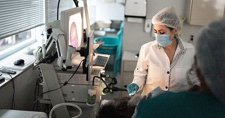
What is Skull Base Surgery: Overview, Benefits, and Expected Results
The new product is a great addition to our lineup.
Our latest product is an exciting addition to our already impressive lineup! With its innovative features and sleek design, it's sure to be a hit with customers. Don't miss out on this amazing opportunity to upgrade your life!
What is Skull Base Surgery? Overview, Benefits, and Expected Results
Skull base surgery is a type of surgery conducted at the base of the skull to treat various conditions affecting the region. It involves accessing the base of the skull to remove lesions caused by tumors, cysts, AVMs, and other conditions. The procedure can be conducted as an endoscopic procedure or as a traditional open procedure.
The surgeons’ goal is to remove the lesion or tumors without causing other damage while retaining functioning anatomy. To accomplish this, surgeons use specialized instrumentation, including precision instruments and endoscopic visualization systems, to ensure minimal damage is done to surrounding tissue.
In some cases, skull base surgery is necessary to provide access to the brain for further treatment or to prepare for a transplant. As such, knowledge of the anatomy of the skull base is essential for successful outcomes.
Overview of Skull Base Surgery
Skull base surgery is a highly specialized type of surgery used to treat diseases or disorders affecting the base of the skull. It is conducted to access the base of the skull and remove lesions, tumors, cysts, aneurysm, and other pathological conditions. It can be done as an open procedure or endoscopic procedure, depending on the complexity of the condition.
Open skull base surgery involves making an incision in a traditional manner or using a retractor system to provide proper exposure. The surgeon will then use precision instruments, endoscopes, and other specialized tools to access and remove the lesion or tumor without damaging nearby tissue or function.
Endoscopic skull base surgery is an advanced type of skull base surgery that uses miniature tools and a tiny camera to access and treat disorders or lesions in the skull base. This technique has been beneficial in treating certain conditions without the need for a large surgical site, allowing patients to recover faster.
Benefits of Skull Base Surgery
Skull base surgery offers several advantages for patients dealing with lesions, tumors, cysts, and other conditions affecting the base of the skull.
Minimally Invasive Procedure
Skull base surgery can be conducted as a minimally invasive procedure using endoscopy. This technique uses small instruments and a tiny camera to access the affected area, reducing the chance of extensive tissue damage and eliminating the need for a large incision.
Reduced Risk of Complications
Skull base surgery is a highly specialized procedure, and it requires an experienced and knowledgeable team of surgeons. This team will use precision tools and techniques to minimize the risk of damaging tissue or nerve pathways while removing the lesion, minimizing the risk of post-operative complications.
Quicker Recovery Time
Open skull base surgery requires a large incision and could take weeks to heal. Endoscopic skull base surgery is minimally invasive and does not require a large incision, allowing the patient to recover quicker with less pain and discomfort.
Expected Results of Skull Base Surgery
Skull base surgery is usually successful in treating lesions, tumors, cysts, and other conditions affecting the base of the skull. The primary goal of the procedure is to remove the lesion or tumor without damaging surrounding tissue or affecting body functions.
The success of the procedure depends on the type of condition being treated, the size of the lesion or tumor, and the experience of the surgeon. The results of the procedure depend on the skill of the surgical team and the condition of the area before the surgery.
If the lesion or tumor is successfully removed, the patient can expect a speedy and full recovery. In cases of cancer, additional treatments, such as radiation or chemotherapy, may be necessary.
Conclusion
Skull base surgery is a highly specialized surgical procedure used to access the base of the skull to remove lesions, tumors, cysts, aneurysms, and other pathological conditions. It can be done as an open procedure or endoscopic procedure, depending on the complexity of the condition. This procedure offers many benefits to the patient, including a faster recovery time and reduced risk of complications. The success of the procedure depends on the type of condition being treated, the size of the lesion or tumor, and the experience of the surgeon.
Definition and Overview
Skull base surgery is a highly specialized set of surgical procedures performed to treat various conditions that affect the base of the skull. This form of surgery is very challenging, dealing with complex cases and lesions that lie in close proximity to vital neurologic structures.
The base of the skull is divided into several compartments, namely the anterior, middle and posterior. Diseases of the anterior fossa typically involve the paranasal sinuses, the orbit, and the nasal cavities. Nasopharyngeal carcinomas (NPCA) are the most frequently encountered tumors in the anterior compartment of the skull base. Masses of the anterior fossa grow slowly; thus, they usually present when they have grown large already, resulting in considerable mass effect and symptoms. Diseases of the middle fossa are usually benign, and include diseases of the pituitary gland and cavernous sinus. Diseases in this area can present with symptoms of cranial nerve involvement, making surgery difficult. Diseases of the posterior fossa involve the cerebellopontine angle and the foramen magnum. Symptoms of posterior fossa diseases include cranial nerve deficits, extremity weakness and gait problems, to name a few.
A multidisciplinary approach is usually necessary when dealing with diseases of the skull base. Neurosurgeons, along with specialists in otorhinolaryngology, ophthalmology and head and neck surgery, primarily manage skull base diseases. Interventional radiologists and oncologists are also involved when dealing with malignant conditions of the skull base. A combination of therapies, including surgery, chemotherapy, radiotherapy and gamma knife treatment, may be needed in the management of various skull base diseases.
Who Should Undergo and Expected Results
Patients with diseases of the skull base are candidates for skull base surgery. These diseases may either be benign or malignant. Benign conditions include congenital abnormalities, vascular malformations, bone diseases, aneurysmal bone cysts and benign tumors such as fibromas and giant cell tumors. Malignant conditions include tumors such as schwannomas (nerve sheath tumors), chondrosarcomas and meningiomas, to name a few.
Imaging studies are essential in the management of skull base diseases. Options include cranial computed tomographic (CT) scan, magnetic resonance imaging (MRI), and cerebral angiography. These studies can help determine the extent of the lesion and can aid in the decision-making regarding surgery and adjuvant therapy. A biopsy of mass lesions may have to be taken to confirm the diagnosis.
If at all possible, the goal of skull base surgery is complete resection of lesions, resulting in resolution of symptoms and functional outcomes. However, not all diseases, particularly tumors in the advanced stages, can be removed entirely. There are several factors to consider prior to subjecting a patient to skull base surgery. These include the patient’s condition and ability to tolerate major surgery, the natural history of the disease, the patient’s symptoms, and the structures that are involved, among others. Patient prognosis depends on the kind and extent of the lesion. Benign lesions can generally be removed with minimal mortality and morbidity.
How Does the Procedure Work?
The goal of skull base surgery is to remove as much of the lesion or mass as possible, with minimal morbidity. The approach to the skull base is mainly dependent on the location of the tumor, and usually involves either a craniotomy or an endoscopic approach. For example, lesions of the anterior skull base may involve an intracranial approach, an extracranial approach, or a combination of both. Middle fossa lesions can be accessed by going through the temporal bone, and diseases of the lateral skull base may be approached via a preauricular or postauricular incision. Posterior approaches, meanwhile, include suboccipital and retromastoid craniotomies. Open approaches involve incisions on the face and scalp, and may necessitate movement of bones. Endoscopic techniques, including endoscopic-assisted operations, allow minimally invasive access to lesions in the brain, and are especially useful when working in areas that are difficult to reach. The endoscope is inserted through the nose, and the disease is approached from below. In recent years, indications for using the endoscopic approach are increasing.
Once access is gained, the mass or the lesion is then removed as completely as possible. The procedure is performed with utmost care using a microscope, in order to preserve the vital structures. The need for reconstruction, such as a free flap, depends on the extent of resection involved.
After the surgery, the patient is transferred to the intensive care unit (ICU). The patient is closely monitored for several days.
Skull base surgery continues to evolve. Significant improvements in this field have been seen, with developments in high-resolution imaging technology, the advent of endoscopic surgery and increasing experience in reconstructive techniques.
Possible Complications and Risk
Skull base surgery is a complex procedure, and various complications can occur during the operation. Neurologic complications are unique to this kind of surgery. Injury to the cranial nerves is not uncommon, and can be caused by electrocautery or traction injury. Direct involvement of the cranial nerves may necessitate complete transection of the cranial nerves. Damage to several cranial nerves can result in devastating consequences, such as difficulty in swallowing, hearing loss, or inability to close the eyes.
Aside from cranial nerve injuries, cerebrospinal fluid (CSF) leaks can also occur. This happens when the dura is invaded by the mass or is opened on purpose. CSF leaks can lead to other complications, such as meningitis. Holes in the dura may be tricky to repair, and may need flaps for adequate coverage.
Other neurologic complications include hydrocephalus, contusion, brain edema, strokes and seizures, to name a few. Significant bleeding is a risk with this kind of operation, as the skull and the meninges are vascular structures. Skull base surgery may also result in scars that are not cosmetically pleasant. It can also produce deformities, such as distorted facial contours, depressions on the scalp, or uneven eyes. Reconstructions using flaps, bone grafts and plates may be necessary to avoid these problems. Complications related to the wound, such as infection, cellulitis or flap necrosis, may also occur.
References:
Kassam A B, Gardner P, Snyderman C H, Mintz A, Carrau R. Expanded endonasal approach: fully endoscopic, completely transnasal approach to the middle third of the clivus, petrous bone, middle cranial fossa, and infratemporal fossa
Snyderman C, Kassam A, Carrau R, Mintz A, Gardner P, Prevedello D M. Acquisition of surgical skills for endonasal skull base surgery: a training program. Laryngoscope. 2007;117:699–705. [PubMed]
/trp_language]
[trp_language language=”ar”][wp_show_posts id=””][/trp_language]
[trp_language language=”fr_FR”][wp_show_posts id=””][/trp_language]


Very informative article! #skullbasesurgery