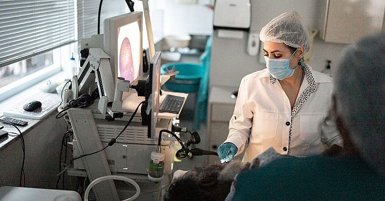
What is Sphincterotomy: Overview, Benefits, and Expected Results
The new product is a great addition to our lineup.
Our latest product is an exciting addition to our already impressive lineup! With its innovative features and sleek design, it's sure to be a hit with customers. Don't miss out on this amazing opportunity to upgrade your life!
What is Sphincterotomy?
Sphincterotomy is a minimally invasive surgical procedure which involves cutting through a muscle, known as the Sphincter, to allow drainage of fluids or excretion of toxins from a body part. It is commonly used to treat bile duct and pancreatic duct obstructions, but it can also be used to address digestive tract distress or other conditions related to muscles, such as anal fissures.
The anatomical structure of the Sphincter can vary depending on the area of the body in which it is located. The most common type of Sphincter is the anal sphincter, which surrounds the lower rectum and anus. Other types of Sphincters are found in the digestive tract, such as the cardiac sphincter and the pyloric sphincter.
Overview of Sphincterotomy
Sphincterotomy is performed under local or regional anesthesia in a surgical center or hospital. During the procedure, a small incision is made through the skin and muscle in the area of the Sphincter. This allows for a physician to view the Sphincter and make small cuts into it for drainage or to correct an obstruction. The incision usually heals without any complications. In some cases, the Sphincter may need to be repaired after the surgery.
Once the Sphincter has been cut, the patient is placed in a recovery room where they are monitored for any potential complications. Depending on the type of procedure, the patient may be able to go home the same day as the surgery or may have to stay in the hospital for a few days for additional monitoring and treatments.
Benefits of Sphincterotomy
Sphincterotomy is a safe and effective treatment for several conditions. It is minimally invasive, meaning that it carries very low risk and requires very little recovery time. Additionally, because it is an outpatient procedure, it is much less expensive than a full abdominal surgery.
In some cases, a Sphincterotomy can relieve symptoms associated with blockages or obstructions. Patients who have had their Sphincterotomy may experience a reduction in pain, bloating, or constipation. It can also reduce the risk of complications and improve overall digestion.
Expected Results of Sphincterotomy
When performed correctly, a Sphincterotomy can improve digestive function and reduce symptoms of digestive issues. It can also help to restore normal levels of fluids and toxins in the body, reduce the risk of infection, and minimize the risk of further blockages or obstructions.
Most patients who undergo a Sphincterotomy experience improved digestion and a reduction in symptoms within days of the procedure. However, some patients may experience a gradual improvement over time as the Sphincter heals.
It is important to note that not everyone who undergoes a Sphincterotomy will experience the same results. In some cases, the Sphincter may not heal properly and may require additional treatments or surgery. Additionally, some patients may experience recurrence of their symptoms after the procedure.
Conclusion
Sphincterotomy is a minimally invasive surgical procedure that is used to improve digestive function and reduce symptoms associated with bile duct and pancreatic duct obstructions. It is a safe and effective treatment that carries minimal risks and requires little recovery time. The expected results of the procedure include improved digestion, reduced symptoms, and reduced risk of infection. While not everyone will experience the same results, most patients do experience improved digestive health and reduced symptoms within days of the procedure.
Definition and Overview
Sphincterotomy is a surgical procedure to repair anal fissure, which is a tear in the anterior or posterior midline of the anus. Although this is the main surgery performed for anal fissure repair, it is normally done only when other non-surgical treatments including laxatives and dietary changes have failed to correct the condition.
The surgery may be either lateral internal, which involves making a small incision in the inner sphincter muscle to decrease the tension and pressure on the anal canal, or advancement anal flap, which uses a graft material from the patient’s own body to provide more blood supply into the tear so it will heal.
Who Should Undergo and Expected Results
Sphincterotomy is performed on patients diagnosed with anal fissure or tear in the anus. This tear may be obvious because of a crack in the skin or due to the presence of a lump next to the fissure. Other common signs and symptoms include bleeding during bowel movements, burning sensation in the anus and its surrounding area, or pain when passing stool.
Although the condition can affect people of all ages, it is commonly diagnosed among pregnant women because of childbirth, children since they tend to strain when they have bowel movements, and those who suffer from constipation. Underlying viral conditions such as HIV or syphilis, which can prevent the fissure from healing, can also cause the condition.
Anal fissures have the ability to heal on their own, so often, doctors treat them with non-surgical options such as stool softeners, laxatives, rest, high-fiber diet, Botox to relax the muscles, pain relievers, and nitroglycerin treatment. However, if the tear does not heal or worsens, especially over the course of six weeks when it is already considered as chronic, the doctor typically recommends surgery.
How Does the Procedure Work?
Anal fissure repair, which is performed by a colorectal surgeon, can be either lateral internal sphincterotomy or advancement anal flap.
In the lateral internal sphincterotomy, the internal sphincter muscle, one of the two muscles that are responsible for controlling the passing of stool, is either stretched or cut to keep the anal canal more relaxed and increase the chances of healing.
The surgery begins by positioning the patient on the operating table in a supine position (face down) while the legs are raised in stirrups or the buttocks placed in a higher position. Local anaesthesia is applied directly to the anal area, although many surgeons prefer general anaesthesia to prevent tear recurrence. The area is also given antiseptic before the surgeon makes a small incision to access the internal sphincter muscle. Using surgical instruments like a scalpel or scissor, the muscle is divided or cut into two or simply stretched. If there is a scar tissue, which indicates a previous fissure, it will also be removed. The incision is then closed up and bandaged.
The advancement anal flap or the dermal flap is often done on patients with chronic fissures by giving the area, which may no longer be healing on its own, with a healthy source of blood supply.
During the surgery, the patient is administered with general anaesthesia. The surgeon then removes a graft material with a healthy blood supply, usually the skin tissue next to the anus, to close the fissure.
Possible Risks and Complications
It is expected that there will be discomfort and pain around an hour or two after the surgery, so the patient is typically given pain relievers and is advised to remain in a supine position to prevent anaesthesia-induced headaches. Infection can also occur, which can be treated with antibiotics, as well as bleeding, which is usually minor and goes away over time.
One of the more serious complications is fecal incontinence or the loss of voluntary control of bowel movement. It is also possible for the fissure to recur even after these surgeries.
Reference
Johnlin FC, Tucker RD, Ferguson S. The effect of guidewires during electrosurgical sphincterotomy.Gastrointest Endosc. 1992. 38:536.
O’Brien JW, Chen SL, Connolly R, Libby ED. Current induction in a fiberglass guidewire compared to conventional wires during simulated papillotomy. Gastrointest Endosc. 1997. 45:493.
/trp_language]
[trp_language language=”ar”][wp_show_posts id=””][/trp_language]
[trp_language language=”fr_FR”][wp_show_posts id=””][/trp_language]


Interesting article! Great read – super helpful!!