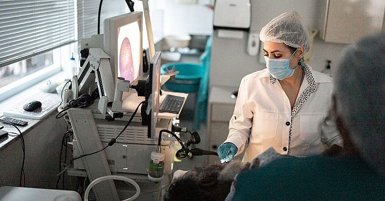
What is Thoracentesis: Overview, Benefits, and Expected Results
The new product is a great addition to our lineup.
Our latest product is an exciting addition to our already impressive lineup! With its innovative features and sleek design, it's sure to be a hit with customers. Don't miss out on this amazing opportunity to upgrade your life!
What is Thoracentesis? Overview, Benefits and Expected Results
Thoracentesis is a medical procedure wherein a doctor or medical professional inserts a needle into the space between the ribcage and lungs known as the pleural cavity in order to access a sample of fluid. The thoracentesis procedure is usually requested for diagnosis and treatment of pleural effusions, a common condition caused by excess fluid in the pleural cavity.
Your doctor may recommend this procedure to diagnose the cause of pleural effusions, relieve the buildup of fluid in the pleural cavity, or remove fluid for further analysis. In some cases, the procedure can be used to deliver medications or chemotherapy drugs directly into the pleural cavity.
How Does Thoracentesis Work
The doctor will perform a physical exam and an imaging scan such as an X-ray or ultrasound to determine the cause of the pleural effusion and the safest area for inserting the needle. The patient will be asked to lie on their side, and the doctor will numb the area of insertion and clean the skin with an antiseptic solution.
A thin needle is inserted between the ribs into the pleural cavity to extract the fluid sample. The doctor will use the imaging scan to guide the needle for maximum accuracy and safety. The amount of fluid removed generally ranges from 10 to 150ml, but more can be collected depending on the size of the fluid buildup.
Benefits of Thoracentesis
Thoracentesis is a fast and effective method of diagnosing and treating pleural effusions. The procedure is considered safe and typically results in a fast recovery without any long-term or serious side effects.
The benefits of thoracentesis include:
- Accurate diagnosis
- Ease and convenience of the procedure
- Effective relief of pleural effusions
- Rapid recovery
- Direct delivery of medication
Expected Results from Thoracentesis
The results of thoracentesis typically depend on the condition for which it was performed. The doctor can collect a fluid sample for further testing to diagnose an infection or determine the cause of the pleural effusions.
If the procedure was performed to remove fluid buildup, the patient can expect immediate relief. Depending on the severity of the fluid buildup, the patient may need to follow up with their doctor for further treatment.
In some cases, the collected sample of fluid can be reinjected into the pleural cavity at a later date. This helps to replenish the fluid in order to reduce symptoms such as pain and shortness of breath.
Are There Any Risks With Thoracentesis?
Thoracentesis is safe for most patients. However, there are some risks associated with the procedure.
Possible Risks Include:
- Infection at the insertion site
- Damage to underlying organs or tissue
- Internal bleeding
- Puncture of the lung
- Collapsed lung
What Should I Expect After Thoracentesis?
The recovery period after thoracentesis depends on the condition for which it was performed. Generally, the patient can expect to experience soreness at the insertion site, which can usually be managed with analgesics such as ibuprofen.
In some cases, the patient may experience drainage or a bandage may need to be applied to the area of insertion. In addition, the patient may need to return to their doctor for follow-up appointments in order to monitor the progression of their condition and the results of the collected sample.
Summary
Thoracentesis is a safe and effective diagnostic and treatment procedure for pleural effusions. The procedure involves the insertion of a needle between the ribs into the pleural cavity to extract a sample of fluid. The benefits of thoracentesis include accurate diagnosis, ease and convenience of the procedure, effective relief of pleural effusions, rapid recovery, and the direct delivery of medication.
Risks associated with the procedure include infection at the insertion site, damage to underlying organs or tissue, internal bleeding, puncture of the lung, and collapsed lung. The recovery period after thoracentesis typically depends on the condition for which it was performed.
Definition and Overview
Thoracentesis is a procedure performed on patients with pleural effusion, which is the build-up of fluid in the pleural space. In a normal individual, there is a balance between continuous fluid production and absorption by the pleura, allowing pleural fluid to be maintained at 10 to 20 ml at any particular time. Any abnormality or disease that affects these processes can lead to the development of pleural effusion.
In thoracentesis, a large bore needle is used to obtain and drain fluid from the pleural space. The pleural fluid samples can then be sent to the laboratory for testing. Pleural fluid examinations allow the analysis of pleural fluid characteristics, identification of bacteria, and determination whether the fluid is a transudate or an exudate. All these tests are useful in determining the etiology of the pleural and in establishing a diagnosis.
Aside from being a diagnostic procedure, thoracentesis can also be therapeutic. The presence of pleural effusion, especially in large amounts, can lead to the compression of the adjacent lung, resulting in symptoms such as difficulty of breathing. Pleural fluid drainage via thoracentesis can lead to remarkable symptom relief in these patients. In some cases, thoracentesis may actually lead to complete resolution of the effusion, with no further intervention necessary.
Who Should Undergo the Procedure and Expected Results
Not all patients with pleural effusion need to undergo thoracentesis. Some patients, particularly those cases wherein the effusion is small, or when it can be attributed to congestive heart failure or uremia, may respond to diuresis or other treatment strategies.
The procedure is recommended:
- In cases wherein the etiology of the pleural effusion is not known
- For patients suffering from pleural effusion for the first time
- For patients with large effusions
- For patients with recurrent pleural effusions
The goal of therapeutic thoracentesis is to remove as much pleural fluid as possible in one sitting. This can produce profound relief in difficulty of breathing. Also, patients with empyema thoracis, or purulent fluid in the pleural space, can benefit from thoracentesis by the removal of the infected fluid.
Adequate drainage of infected effusions and source control are cornerstones in the management of empyema. Finally, drainage of pleural effusion allows a clearer radiographic evaluation of the lungs, which can aid in the proper management of the disease.
How Does the Procedure Work?
The procedure is performed with the patient typically in the sitting or upright position. In bedridden patients where sitting is not possible, putting them in the lateral decubitus position, where they lie down on the side with the effusion, is an alternative. Once properly positioned, the area for thoracentesis is determined. Percussion and auscultation of the chest, along with an imaging study, can help in selecting the best point of entry for the bore needle. The procedure can be performed blindly, with the needle inserted 2 to 3 cm below the most superior border of the effusion, usually along the patient’s back. Smaller or loculated effusions, however, are best drained with ultrasound guidance.
The area is then exposed and cleaned. The thoracentesis needle, usually gauge 18 or 20, is then inserted right above the superior aspect of the rib. This ensures that the intercostal bundle, containing the nerve and vessels, are avoided. The needle is inserted up to the parietal pleura, and pleural fluid is then aspirated. If, instead of fluid, air is aspirated, the thoracentesis site may be too high. Unsuccessful thoracentesis may be due to several reasons, such as the pleural effusion being too viscous or the chest wall being too thick. Using longer and larger bore needles may help, as well as performing the procedure under ultrasound guidance.
Depending on the kit used, the needle may be connected to a 3-way stopcock, which allows multiple aspirations using a syringe, without opening the pleural cavity to air. Some kits allow the insertion of a catheter over a wire that can be inserted through the needle to reach the pleura. Pleural fluid is then drained, and samples are placed in sterile containers for examination. Once the procedure has been completed, the needle or catheter is removed, and the site for thoracentesis is covered with a sterile dressing.
Possible Complications and Risks
Although thoracentesis is a simple procedure, it is not without complications. The most common complication encountered is pneumothorax, or air in the pleural space. Previous studies have reported the incidence of post-thoracentesis pneumothorax to be 11%. Of these, 2% of patients required further management with a tube thoracostomy. Most cases can be managed conservatively, with high oxygen support.
Bleeding is another possible risk of the procedure. When performed properly, bleeding is uncommon. However, this can occur in elderly patients who have tortuous intercostal vessels. It is also a risk in patients with coagulation abnormalities who develop pleural effusions. This risk can be minimized by using a smaller needle when performing the procedure.
A rare, but dreaded complication of thoracentesis is the re-expansion pulmonary edema. This condition is typically caused by the rapid expansion of a collapsed lung. Previously, it was believed that this is associated with the removal of large amounts of pleural fluid; although no consensus exists as to how much fluid can be safely removed, experts recommend a maximum of 1 Li of pleural fluid drainage at any one time. Recent studies, however, are implicating that more than the amount of effusion, it is the speed of drainage that induces the development of this condition.
Other uncommon complications include empyema caused by the introduction of bacteria into the pleural cavity, and tumor spread over the tract of the needle.
References:
Broaddus C, Light RW. Pleural effusion. In: Mason RJ, Broaddus CV, Martin TR, et al, eds. Textbook of Respiratory Medicine. 5th ed. Philadelphia, PA: Saunders Elsevier; 2010:chap 73.
Celli BR. Diseases of the diaphragm, chest wall, pleura, and mediastinum. In: Goldman L, Schafer AI, eds. Goldman’s Cecil Medicine. 24th ed. Philadelphia, PA: Saunders Elsevier; 2011:chap 99.
/trp_language]
[trp_language language=”ar”][wp_show_posts id=””][/trp_language]
[trp_language language=”fr_FR”][wp_show_posts id=””][/trp_language]


Great overview! #Thoracentesis
Nicely detailed!
Great overview! #Thoracentesis Nicely detailed! #superhelpful