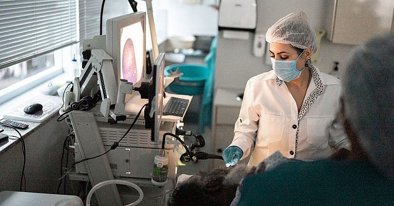
What is Ventriculostomy: Overview, Benefits, and Expected Results
The new product is a great addition to our lineup.
Our latest product is an exciting addition to our already impressive lineup! With its innovative features and sleek design, it's sure to be a hit with customers. Don't miss out on this amazing opportunity to upgrade your life!
What Is Ventriculostomy?
Ventriculostomy is a surgical procedure in which a catheter is placed into a cerebral ventricle to allow for the collection and drainage of cerebrospinal fluid (CSF). It is used to diagnose and treat a variety of medical conditions, such as hydrocephalus, pseudotumor cerebri, subdural hematoma, and brain tumors.
Overview
Ventriculostomy is a procedure during which a catheter is introduced through a tiny hole in the skull. The hole is made at the desired location close to one of the four cerebral ventricles, and the catheter is inserted into the ventricular system. After the catheter placement, a syringe is attached to the catheter, and CSF can be withdrawn from the ventricle.
During the procedure, CSF samples are also obtained for diagnostic purposes. The CSF is then sent for analysis if needed. Ventriculostomy can also be used for therapeutic purposes, where a solution is injected directly into the ventricle in order to reduce intracranial pressure or to provide relief to the patient.
Benefits
Ventriculostomy has several advantages over other forms of cerebral treatment. It is less invasive than other treatments, such as open brain surgery, and poses less risk to the patient. It is also safer and more precise than other forms of diagnostic tests, such as spinal taps, because the catheter can be placed directly into the brain ventricle.
This procedure provides the physician with insight into the condition of the patient by allowing direct access to the CSF. It can also be used to diagnose a variety of medical conditions in the brain by examining the CSF. Ventriculostomy is also useful for therapeutic purposes, such as injecting medication directly into the brain, or for draining excess CSF in patients with hydrocephalus.
Expected Results
The results of the ventriculostomy procedure are highly dependent on the medical conditions being treated. Generally, ventriculostomy can help diagnose and manage long-term cerebral problems such as hydrocephalus and pseudotumor cerebri. It can also help reduce headache and pressure in the brain if the procedure is done for therapeutic purposes.
In cases of hydrocephalus, the procedure is performed in order to reduce intracranial pressure, and is sometimes accompanied by the insertion of a shunt. A shunt is a device that is placed in the brain in order to allow for the continuous drainage of excess CSF.
Ventriculostomy can also be used to diagnose brain tumors, and if it is found that a tumor is present, additional treatment may be required.
Risks and Complications
Ventriculostomy is generally a safe procedure, however, as with any surgical procedure, there are some risks and complications associated with this type of procedure. These include the risks of infection and bleeding, damage to the brain, stroke, and the risk of the catheter migrating away from the intended brain ventricle.
In rare cases, the procedure can lead to an expansion of the ventricles which can cause further intracranial pressure. It is important for the patient to be closely monitored during and after the procedure in order to reduce the risk of complications.
Conclusion
Ventriculostomy is a procedure in which a catheter is placed into one of the cerebral ventricles to collect and drain cerebrospinal fluid (CSF). This procedure is used to diagnose and treat many medical conditions, such as hydrocephalus, pseudotumor cerebri, subdural hematoma, and brain tumors. The procedure is generally safe, with risks of infection, bleeding, damage to the brain, stroke, and the catheter migrating away from the intended brain ventricle.
On the whole, ventriculostomy is a safe and effective procedure that can be used to diagnose and/or treat a variety of medical conditions in the brain. It is important to discuss the risks and benefits with your doctor in order to determine if this procedure is right for you.
Definition and Overview
Ventriculostomy or ventricular drain is a quick surgical procedure performed in the head to attach a device to drain cerebrospinal fluid (CSF) buildup in the brain. This device may be placed externally, and it can be either temporary or permanent.
The procedure is done on the ventricles of the brain, a group of cavities where CSF accumulates. For a number of reasons, they can be obstructed or contain an excessive amount of CSF that increases intracranial pressure. The device often stays attached until cranial pressure normalises.
Who Should Undergo and Expected Results
Ventriculostomy is recommended for patients when there’s CSF buildup in the brain. CSF is a transparent fluid found in the central nervous system (brain and spine) that provides additional protection for the brain’s cortex. After the CSF is released, it should be reabsorbed into the bloodstream to help maintain the ideal cranial pressure.
There are many reasons why CSF accumulates in the brain ventricles. These include hydrocephalus, a general term used for conditions wherein the spaces in between the ventricles widen, leaving less room for CSF to flow or be reabsorbed. A landmark symptom is the prominent big head.
Hydrocephalus may be acquired or genetic. The latter occurs during foetal development while the former means that it developed after the baby is born. A specific procedure performed to treat it is called endoscopic third ventriculostomy.
Intracranial pressure can also be due to a traumatic brain injury. This is considered a medical emergency as the pressure can increase rapidly, increasing the risk of permanently damaging the brain tissues.
In other cases, ventriculostomy is performed to deliver the medications to treat brain ventricle or other brain-related problems and to diagnose CSF, which may indicate the presence of infection or other underlying conditions causing neurological symptoms.
It’s expected that as soon as the ventricular drain is added, brain pressure would go down and normalise.
How Does the Procedure Work?
Before the procedure is performed, a neurosurgeon conducts a pre-surgical consultation, wherein he assesses the patient’s overall neurological and physical health. If the patient is taking certain medications, he may have to stop some of them, especially if they can trigger blood loss or clotting during and after the operation.
Usually, the procedure lasts for about an hour and general anaesthesia is typically not a requirement. Only local anaesthesia is applied to the part of the scalp where an entry point will be made. The patient can also be given sedation for maximum comfort.
During the procedure, the patient lies on his back on the operating table. A part of the scalp is shaved and cleaned while the rest of the head is covered with a surgical drape. The surgeon then proceeds to create a hole in the head using a surgical drill to access the dura mater. In some cases, this is enough to improve cranial pressure. However, if ventricular drain should be continued, a tube is attached directly to the ventricle, and a bag is connected to the tube. The patient is also connected to a system that monitors the level of CSF and brain pressure.
If the tube is only temporary, it’s called an EVD (external ventricular drain). If it’s permanent, it’s called a shunt. The tube is kept in place with sutures while the drain stays in place.
Risks and Complications
One of the possible complications of the procedure is infection, which happens because of the wound site. If this infection is left untreated, it may result in sepsis or the systemic inflammation of the body that can cause multiple organ failure. There may also be bleeding. Patients are advised not to touch the bag while it’s still draining.
It’s also possible that the bag will leak or that too much of CSF is drained, a condition known as hypotension, which can cause serious headaches.
Reference:
- Rosenberg GA. Brain edema and disorders of cerebrospinal fluid circulation. In: Daroff RB, Fenichel GM, Jankovic J, Mazziotta JC, eds. Bradley’s Neurology in Clinical Practice. 6th ed. Philadelphia, Pa: Saunders Elsevier; 2012:chap 59.
/trp_language]
[trp_language language=”ar”][wp_show_posts id=””][/trp_language]
[trp_language language=”fr_FR”][wp_show_posts id=””][/trp_language]


Interesting read! #ventriculostomy
Great source of info! #ventriculostomy
#ventriculostomy Very informative!