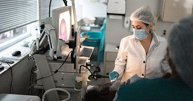
What is Submandibular Gland Excision: Overview, Benefits, and Expected Results
The new product is a great addition to our lineup.
Our latest product is an exciting addition to our already impressive lineup! With its innovative features and sleek design, it's sure to be a hit with customers. Don't miss out on this amazing opportunity to upgrade your life!
What is Submandibular Gland Excision?
Submandibular gland excision, also known as submandibular salivary gland resection, is a procedure used to remove a salivary gland located near the lower jaw. This surgical operation is often performed to reduce the amount of saliva being produced and to treat certain types of tumors in the submandibular region.
Typically, the submandibular glands are located on either side of the mouth, just beneath the lower jawbone. The excision of these glands can be done through either a traditional open surgical procedure or through a minimally-invasive laparoscopic technique. In either case, the procedure requires the use of general anesthesia to perform.
Overview of Submandibular Gland Excision
During a submandibular gland excision, a surgeon will make a small incision in the lower face near the submandibular gland. The entire gland will then be removed along with some surrounding tissue. The wound will be closed with either sutures or staples, and a bandage will be applied.
In a traditional open submandibular gland excision, the patient will typically spend several days in the hospital following the procedure for recovery and observation. In the case of minimally-invasive laparoscopic submandibular gland excision, most patients can return home the same day.
Benefits of Submandibular Gland Excision
There are a few key benefits associated with submandibular gland excision.
- Reduced saliva production: By removing the salivary gland, the body’s production of saliva will be reduced. This can help reduce excessive drooling, which can be a common problem among those with multiple sclerosis or head and neck cancer.
- Reduced risk of tumors: Removing the salivary gland can help reduce the risk of tumors developing in the submandibular region.
- Improved quality of life: For those with conditions that cause excessive drooling, submandibular gland excision can significantly improve their quality of life.
Expected Results
The expected results following a submandibular gland excision will vary depending on the patient and the underlying condition. However, the most common result is the reduced production of saliva. This can significantly reduce or eliminate the symptoms of excessive drooling.
It is also important to note that patients may experience some difficulty in chewing or swallowing for the first week or two following the procedure. However, once the area has healed most of these side effects should quickly dissipate.
Risks and Complications
As with any surgery, there are a few risks and complications associated with submandibular gland excision. These include bleeding, infection, and abnormal scarring or tissue changes. Other potential complications include nerve damage, which can result in problems with chewing, swallowing, or speaking. Additionally, there is a slight risk of developing a salivary fistula, which is an abnormal connection between the salivary duct and a neighboring structure.
Preparing for Submandibular Gland Excision
Before a submandibular gland excision is performed, the surgeon will typically order tests such as CT scans or X-rays to get a better look at the salivary glands. Additionally, the patient will need to discuss any medications, supplements, or herbal remedies they are taking with the surgeon.
Conclusion
Submandibular gland excision is a surgical operation used to remove a salivary gland located near the lower jawbone. This procedure can help reduce the body’s production of saliva and reduce the risk of developing tumors in the submandibular region. While there are some risks associated with the procedure, it is typically safe and successful.
Before undergoing submandibular gland excision, it is important to discuss any medications, supplements, or herbal remedies that are being taken with the surgeon. Additionally, tests such as CT scans or X-rays may be ordered to get a better look at the gland before the operation.
Definition and Overview
There are three major salivary glands in the body: the parotid, submandibular, and sublingual glands. Just like any other part of the body, these glands can also develop tumours. The majority of salivary gland tumours are found in the parotid gland but most of them are benign. Meanwhile, half of the masses discovered in the submandibular and sublingual glands turn out to be malignant. Management of these masses entails the removal of the gland via surgery.
Who Should Undergo and Expected Results
Tumour excision is an important aspect in the management of patients with masses of the submandibular gland. Malignant tumours of the submandibular gland may range from low to high grade, and activity depends on the kind of tumour and the stage. Commonly encountered submandibular gland tumours are adenoid cystic carcinoma and mucoepidermoid carcinoma.
Tumours of the submandibular gland usually manifest as a palpable mass on the neck. Pain may also be an associated symptom as well as invasion of the nerves, particularly the hypoglossal nerve, which may result in paralysis.
Low-grade tumours generally have a good prognosis, with 5-year survival rates reaching up to 70%. High-grade tumours, on the other hand, do not have as good a prognosis. In certain cases that involve nerve invasion and regional metastases, radiation therapy may be indicated postoperatively.
Another indication for submandibular gland excision is the chronic inflammation of the gland due to the formation of stones. Rarely, patients who experience trauma to the submandibular area may also require excision of the submandibular gland.
How is the Procedure Performed?
Submandibular gland excision is performed with the patient asleep under general anaesthesia, with the head rotated to the side opposite the tumour. The basic principle employed in this procedure is en bloc removal of the entire gland, including the submandibular lymph nodes, with preservation of nerves, if possible.
Operations in the submandibular area begin with a curvilinear incision along the mandible, starting from the midline to the mastoid process (near the ear). A flap is then created under the platysma muscle, which remains attached to the skin. The facial nerve, specifically its marginal branch, is then identified, located immediately underneath the muscle. The nerve should be preserved unless the tumour has also invaded it.
The gland is then gently dissected, beginning at the level of the hyoid bone. The hypoglossal nerve is identified, which is located between the digastric muscle and the gland itself. The lingual nerve is likewise identified, which can be found upon the retraction of the mylohyoid muscle.
Dissection is continued up to the facial artery, which serves as the blood supply to the submandibular gland. The artery is ligated, making sure to have an adequate margin just in case the artery retracts under the digastric muscle. The excision of the submandibular gland is completed with the ligation of the Wharton duct.
Some surgeons send the specimen for frozen section biopsy to determine if the resected margins are adequate. The specimen is examined while the patient is still in the operating theatre, and a decision is made based on the histologic findings. More extensive resections may be required if other adjacent structures, such as the mandible, are affected. Meanwhile, the presence of enlarged lymph nodes in the neck may require further neck dissection during the same operation.
Finally, a drain is inserted and closure is performed in layers. A sterile dressing is applied to the wound afterwards.
Possible Risks and Complications
The excision of the submandibular gland requires a thorough knowledge of head and neck anatomy, as a number of important structures can be found in this area. Injuries to these vital structures can result in significant and permanent morbidity.
One of the most serious and disabling complications of the procedure is an injury to the nerves, particularly the facial nerve, the hypoglossal nerve or the lingual nerve. Approximately 10-30% of cases end up with facial paralysis or paresis after the operation. Most of these cases are temporary and are due to the stretching of the facial nerve during dissection or to inflammation after the operation and usually resolve within a few weeks to months. However, 7-12% of these cases result in permanent facial paralysis, leading to the inability to move the muscles of the face near the lips. Permanent damage to the lingual and hypoglossal nerves occur less frequently (2-5%). Occasionally, the nerves need to be sacrificed due to direct involvement with the tumour. In these cases, immediate nerve grafting during the operation may be necessary.
Aside from nerve injury, other complications of the procedure include infection and wound-related problems, such as scar formation. Bleeding and hematoma formation can be minimised with proper hemostasis while the application of compressive dressings and use of drains minimise the risk of seroma formation. Tumour recurrence is also a possibility, and may be associated with inadequate excision or margins of resection. The development of salivary fistulae has also been reported. In some benign conditions, chronic inflammation of the area may occur due to residual stones in the salivary ducts.
References
Strympl P, Kodaj M, Bakaj T, Kominek P, Starek I, Sisola I, et al. Color Doppler Ultrasound in the pre-histological determination of the biological character of major salivary gland tumors. Biomed Pap Med Fac Univ Palacky Olomouc Czech Repub. 2012 Sep 5.
Witt BL, Schmidt RL. Ultrasound-guided core needle biopsy of salivary gland lesions: a systematic review and meta-analysis. Laryngoscope. 2014 Mar. 124 (3):695-700.
/trp_language]
[trp_language language=”ar”][wp_show_posts id=””][/trp_language]
[trp_language language=”fr_FR”][wp_show_posts id=””][/trp_language]


So informative! #medicine Great resource for learning about the procedure and its benefits!
I’m so glad my dentist suggested this procedure
#SoHelpful – Thank you for sharing this overview of Submandibular Gland Excision – I’m sure many people will benefit from reading it!