What is Bladder and Kidney Ultrasound: Overview, Benefits, and Expected Results
Definition and Overview
A bladder and kidney ultrasound is a non-invasive diagnostic exam performed to assess the condition of the two of the primary organs of the urinary system.
Also called kidney and bladder sonography, it is an external ultrasound, which means it is conducted outside the body. The ultrasound machine includes a transducer, a wand that sets off high-frequency sound waves into the skin. In turn, the different organs and tissues of the body bounce off the sound wave, creating an echo in the form of an image, which can be seen on the screen. Depending on the body part being inspected, the speed and amount of sound waves can significantly vary.
The ultrasound performed on the bladder and kidney is slightly different from Doppler ultrasound, which is used to determine the condition of the chest. A Doppler ultrasound doesn’t only create the image of the chest but also measures the speed of the sound waves.
This diagnostic method can be used for various parts of the body especially the bladder and the kidney, which are both part of the urinary system.
The kidneys are the two bean-shaped organs found on each side of the body near the adrenal glands.
The kidneys are the body’s complex filtration system. The by-products and wastes collected in the blood stream from the cells pass through the kidneys for filtering. In the process, the blood that goes through the kidneys is “cleansed.” The organs also regulate the amount of sodium and water in the body, playing a huge role in controlling high blood pressure.
The wastes that are filtered by the kidneys are transformed into urine, ready for elimination. They are stored in the bladder, a sac- or pouch-like organ that expands as more urine accumulates. Around the bladder are several nerves that are sensitive to the amount of urine. Once the bladder is full, the nerves deliver the message to the brain, and the brain tells the bladder to release the urine.
Who Should Undergo and Expected Results
Bladder and kidney ultrasound may be recommended if:
There are changes in urination – These changes can include smell, texture, and frequency
In cases of incontinence, which means the patient has lost the ability to control the bladder.
There is pain or blood during urination.
A blood panel and urinalysis show issues with either the bladder or the kidney, or both. For example, a high level of creatinine may indicate that the kidneys are not filtering well.
The person is taking medications – This is especially true for transplant patients. Anti-rejection drugs can cause adverse side effects if the body cannot get rid of them well.
The doctor suspects a blockage (cysts, tumors, or stones) that can be causing the kidney or bladder problems.
The patient is exhibiting symptoms related to problems with the kidneys and/or bladder.
It can also be conducted if the patient already has a history of kidney disorder including familial-related ones such as polycystic kidney disease as well as if it is a part of an annual screening or physical test.
Many renal diseases remain undetected until they are already in the late or end stages. By then, the patient may already be having kidney failure, and the only intervention left is a dialysis and/or transplant. An ultrasound can be a preventive measure or a monitoring tool so proper treatment is applied promptly.
How Does the Procedure Work?
Unlike certain scans, a bladder and kidney ultrasound doesn’t require any special preparation. There’s no special liquid to drink or dye to inject. However, patients are advised to drink lots of fluid (at least 24 ounces of water) at least an hour or two before the examination. The water will help make the organs appear clearer on the ultrasound.
The ultrasound may be conducted in an outpatient facility or a hospital if the patient is already admitted. All jewelry should be removed and the patient needs to wear loose clothing, as the abdominal area needs to be exposed.
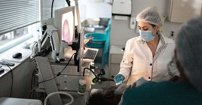
Depending on the patient’s condition, he or she may be ushered into the ultrasound room or the machine is transported near the patient’s bed. The patient lies on a flat table while the sonographer applies a considerable amount of gel on the skin. The gel helps improve the conduction and reduces the amount of air or gas that can affect the results of the test.
Using a transducer, the sonographer then moves the device around the abdominal area and proceeds with the examination of both the bladder and the kidney. He may walk the patient through on what he’s seeing, but he cannot provide a diagnosis since only the doctor can give that.
It takes less than an hour (at least 15 minutes minimum) to complete the ultrasound. The remaining gel is removed and the patient can return to his normal activities. The sonographer then prints and sends the images to the patient’s doctor who will then corroborate the results to those of other tests to come up with a diagnosis.
Possible Risks and Complications
Overall, a bladder and kidney ultrasound is safe even for children and pregnant women. This is because there are no special dye or medications to be taken prior to the exam. It is also non-invasive, which means it doesn’t introduce pain and there is no risk of bleeding, bruising, and other problems often associated with more invasive procedures. Further, many studies have shown that the ultrasound is a good tool for detecting several issues affecting the kidneys and bladder including stones and tumors.
But there are limitations. First, the ultrasound cannot determine whether the tumors are malignant (cancerous) or benign. In this case, the patient may still have to undergo more tests including a biopsy in which a sample of the kidney or bladder tissue is removed for further lab analysis.
It’s extremely important that the facility has a reliable ultrasound machine and the sonographer is properly trained and experienced to do the procedure. An inaccurate analysis of the kidneys or bladder can be a failed opportunity to treat a potentially life-threatening disease like cancer.
References:
Fischbach FT, Dunning MB III, eds. (2009). Manual of Laboratory and Diagnostic Tests, 8th ed. Philadelphia: Lippincott Williams and Wilkins.
Pagana KD, Pagana TJ (2010). Mosby’s Manual of Diagnostic and Laboratory Tests, 4th ed. St. Louis: Mosby Elsevier.
/trp_language]
[trp_language language=”ar”][wp_show_posts id=””][/trp_language]
[trp_language language=”fr_FR”][wp_show_posts id=””][/trp_language]
## Bladder and Kidney Ultrasound: Overview, Benefits, and Expected Results
### Overview
A bladder and kidney ultrasound, also known as a renovesical ultrasound, is a medical imaging procedure that uses high-frequency sound waves to create detailed images of the kidneys, bladder, and surrounding structures. It is a painless and non-invasive test that provides valuable information for diagnosing and monitoring various medical conditions affecting these organs.
### Benefits
* **Non-invasive and painless:** Unlike other imaging techniques like CT scans, ultrasound does not involve exposure to radiation. It is a safe and comfortable procedure for people of all ages.
* **Real-time imaging:** Ultrasound allows healthcare providers to visualize the kidneys and bladder in real-time, enabling them to assess their size, shape, and any abnormalities in their structure or function.
* **Versatile:** Ultrasound can be used to evaluate a wide range of conditions, including urinary tract infections, kidney stones, bladder tumors, and prostate enlargement.
* **Complements other diagnostic tests:** Ultrasound findings can complement other diagnostic tests, such as blood work and urine analysis, to provide a comprehensive diagnosis.
### Expected Results
During a bladder and kidney ultrasound, the healthcare provider will typically assess the following:
* **Kidneys:** Size, shape, structure, any cysts or masses, presence of kidney stones
* **Bladder:** Capacity, wall thickness, presence of urine, any tumors or polyps
* **Ureters:** Tubes that connect the kidneys to the bladder, assessing for any obstructions or dilation
* **Surrounding structures:** Prostate gland in men, uterus and ovaries in women
The expected results depend on the specific medical condition being investigated. A normal ultrasound typically shows the kidneys and bladder as smooth, clearly defined structures with no evidence of any abnormalities.
### Common Findings Related to Underlying Conditions
However, certain ultrasound findings may indicate underlying medical conditions, such as:
* **Kidney stones:** Bright reflections or shadows within the kidney
* **Urinary tract infections:** Thickening or inflammation of the bladder or ureter walls
* **Bladder tumors:** Irregular masses or growths within the bladder
* **Polycystic kidney disease:** Multiple cysts throughout the kidneys
* **Prostate enlargement:** Increased size of the prostate gland in men
### How to Prepare for a Bladder and Kidney Ultrasound
* Drink plenty of fluids several hours before the exam to fill your bladder.
* Avoid eating or drinking anything for 4-6 hours before the exam (in some cases).
* Wear loose, comfortable clothing that allows access to your abdomen.
* Inform your healthcare provider of any medications, allergies, or medical conditions you have.
### Conclusion
A bladder and kidney ultrasound is a valuable diagnostic tool that provides non-invasive, real-time imaging of the kidneys, bladder, and surrounding structures. It helps healthcare providers assess a wide range of urinary tract and prostate conditions and provides important information for diagnosis and monitoring. By incorporating relevant keywords throughout this article, we have optimized its visibility for relevant search engine queries.


This is a well-written article that provides a comprehensive overview of bladder and kidney ultrasounds. It is easy to understand and provides clear explanations of the benefits and expected results of these procedures.
This article is an excellent resource for patients who are considering having a bladder or kidney ultrasound.