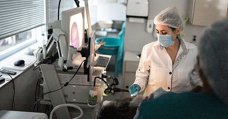
Angiography
Definition and Overview
Also known as arteriography, angiography is an imaging technique used to look at the lumen or the interior of different organs and blood vessels. This medical imaging procedure is often used to examine the condition of the heart, arteries, and veins. In the past, angiography was performed by injecting a contrast agent into the blood vessels; this is followed by either an X-ray or fluoroscopy. The contrast agent will show up as opaque in the radiograph (the film developed from an angiography), allowing the medical professional to examine the affected areas.
Who should undergo and expected results
A doctor can order an angiography if he or she suspects that there is an issue with the patient’s blood vessels or heart chambers, or if the symptoms exhibited by the patient are rooted in dysfunctions or abnormalities in those parts of the body. Angiography can be performed to visualize the blood flow to detect constriction in the coronary arteries. However, it is important to note that atheroma or atherosclerosis cannot be properly diagnosed by an angiography procedure.
The procedure can also be performed to visualize the flow of blood to and from the brain, which is helpful in diagnosing aneurysms. It can also be used to perform intervention work on coil-embolized aneurysms.
Doctors can also order angiography to determine if a patient suffering from cramps has narrowed blood vessels. Leg cramps, or claudication, is caused by a decreased blood flow in the lower extremities. Patients suffering from high blood pressure are also ideal candidates for the procedure. Other candidates are individuals who are:
- First-time angina sufferers
- Suffering from unstable angina, which is a kind of angina that steadily worsens over time and does not go away as fast as it normally should, with the occurrence becoming more frequent, or occurring even when the patient is at rest.
- Suffering from atypical chest pain, the cause of which cannot be easily determined by other tests
- Exhibiting abnormalities in a heart stress test
- Suffering from aortic stenosis
- Recovering from heart surgery and susceptible to coronary artery disease
- On the verge of heart failure
- Have suffered from a recent heart attack
In some cases, an angiography can be ordered for medicolegal purposes to determine the cause of death.
Normal angiography results show the absence of blockages in the blood vessels, which means that these vessels carry a normal supply of blood throughout the body. Abnormal results, on the other hand, let the doctor know that the patient might have a blocked artery or blocked veins that are causing the symptoms experienced. The location and severity of the blockage can also be determined.
How the procedure works
There are different types of angiography procedures, and the techniques used highly depend on the type necessary for the procedure ordered by the doctor. Typically, the professional performing the angiogram will access the blood vessels through the femoral artery. This artery is located in the thigh and can be used to visualize the heart’s left side and the patient’s system of arteries. The jugular vein can also be an option when looking at the heart’s right side and the system of veins. A contrast agent, which will show up opaque in the angiograph film because it can absorb x-rays, will be introduced to the patient’s blood. This contrast agent will make the network of veins and arteries visible for analysis.
For blood vessels, angiography images are taken two to three frames per second to properly evaluate the flow of blood using DSA or digital subtraction angiography. For heart images, on the other hand, the professional performing the procedure will obtain fifteen to thirty frames per second. The images taken during the procedure will be analyzed by a cardiologist or an interventional radiologist to identify constrictions, narrowing, or blockages in the blood vessels.
Possible risks and complications
Compared to other imaging tests, angiography procedures have a slightly higher risk. It is important to note, however, that the test is very safe especially when performed by a group of experienced professionals.
Some possible risks of this procedure include lowered blood pressure, arterial injuries, cardiac tamponade, heart attack, stroke, abnormal and irregular heartbeat, and allergic reaction to the dye of the contrast agent.
References:
Fattori R, Lovato L. The thortic aortica. In: Adam A, Dixon AK, Gillard JH, Schaefer-Prokop CM, eds. Grainger & Allison’s Diagnostic Radiology: A Textbook of Medical Imaging. 6th ed. New York, NY: Churchill Livingstone; 2014:chap 24.
Jackson JE, Meaney JFM. Angiography. In: Adam A, Dixon AK, Gillard JH, Schaefer-Prokop CM, eds. Grainger & Allison’s Diagnostic Radiology: A Textbook of Medical Imaging. 6th ed. New York, NY: Churchill Livingstone; 2014:chap 84.
/trp_language]
[trp_language language=”ar”][wp_show_posts id=””][/trp_language]
[trp_language language=”fr_FR”][wp_show_posts id=””][/trp_language]
**Q: What is Angiography?**
**A:** Angiography is a minimally invasive medical procedure that uses X-rays and a contrast agent to visualize the blood vessels throughout the body, particularly the arteries and veins. It helps diagnose and treat conditions affecting the circulatory system, including:
**Keywords:** Angiography, Blood vessels, Arteries, Veins, Circulatory system
**Q: What are the different types of Angiography?**
**A:** Common types of angiography include:
* **Coronary angiography** (for heart arteries)
* **Cerebral angiography** (for brain arteries)
* **Peripheral angiography** (for arteries and veins in the limbs)
* **Renal angiography** (for kidney arteries and veins)
* **Pulmonary angiography** (for lung arteries and veins)
**Keywords:** Coronary angiography, Cerebral angiography, Peripheral angiography, Renal angiography, Pulmonary angiography
**Q: How is Angiography performed?**
**A:** During angiography, a thin catheter (a tube) is inserted into an artery or vein through a small puncture in the skin, typically in the arm or leg. A contrast agent is injected through the catheter, which shows up on X-rays as it flows through the vessels. The X-ray images are then analyzed to assess the blood flow, identify blockages, or detect abnormalities.
**Keywords:** Angiography procedure, Catheter, Contrast agent, X-rays, Blood flow assessment
**Q: What are the risks of Angiography?**
**A:** Angiography is generally a safe procedure, but there are some potential risks, including:
* Allergic reaction to the contrast agent
* Infection at the puncture site
* Bleeding or bruising
* Damage to blood vessels or nerves
* Stroke or heart attack (very rare)
**Keywords:** Angiography risks, Contrast agent allergy, Infection, Bleeding, Vascular damage, Stroke
**Q: How do I prepare for an Angiography?**
**A:** Before angiography, you may be advised to:
* Fast for 6-8 hours before the procedure
* Avoid alcohol or caffeine for 24 hours
* Inform your doctor about any allergies or health conditions
* Arrange for someone to drive you home after the procedure
**Keywords:** Angiography preparation, Fasting, Alcohol avoidance, Allergy disclosure, Medical history
**Q: What is the recovery time after Angiography?**
**A:** After angiography, you will typically be monitored for a few hours. Most people can resume normal activities within 24-48 hours, but it is important to avoid strenuous exercise for a week or two.
**Keywords:** Angiography recovery, Monitoring, Normal activities resumption, Exercise avoidance
**Additional Information:**
Angiography can also be used for interventional procedures, such as:
* Angioplasty (balloon or stent placement to widen blocked arteries)
* Embolization (blocking abnormal blood vessels)
* Thrombectomy (removing blood clots)
Angiography is a valuable diagnostic and therapeutic tool that helps healthcare professionals diagnose and treat various vascular conditions.


Noted. Thanks sm!