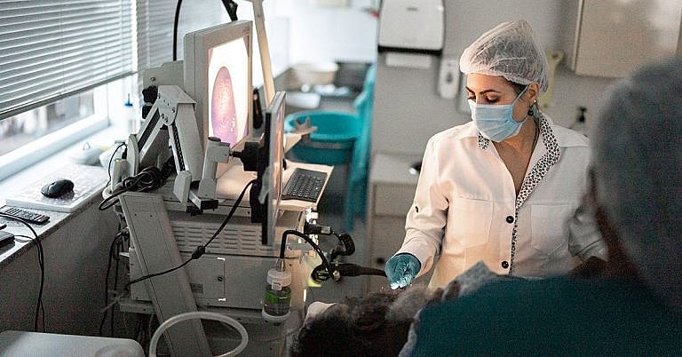
What is Thoracoscopy: Overview, Benefits, and Expected Results
The new product is a great addition to our lineup.
Our latest product is an exciting addition to our already impressive lineup! With its innovative features and cutting-edge design, it's sure to be a hit with customers. Don't miss out on this amazing opportunity to upgrade your life!
What is Thoracoscopy: Overview, Benefits, and Expected Results
Thoracoscopy is a minimally invasive surgical procedure that uses a video camera to get an inside view of the chest cavity. It’s used to diagnose and treat conditions such as pleural diseases, lung nodules, pneumothorax, and tumors. It can also be used to remove fluid buildup in the chest cavity, diagnose internal bleeding, and evaluate the lungs and chest wall during biopsies.
How Does Thoracoscopy Work?
Thoracoscopy is performed under general anesthesia. An incision is made in the chest cavity, through which the thoracoscope (a thin, lighted tube) is inserted. The thoracoscope is connected to a video camera, which provides a live video feed of the inside of the chest cavity to a monitor that the doctor can observe. The video feed allows the doctor to examine the organs and structures inside the chest without having to make a larger incision.
What Are the Benefits of Thoracoscopy?
Thoracoscopy offers several benefits over traditional open surgery, including:
- Small incisions: Thoracoscopy requires only a few small incisions. This means less pain and faster healing times.
- Less risk of infection: Because the incisions are much smaller, there’s less risk of infection.
- Shorter hospital stays: Thoracoscopy is associated with shorter hospital stays and faster recovery times.
- Less scarring: The small incisions used for thoracoscopy result in less scarring than traditional open surgeries.
- Improved precision: The video camera used in the procedure helps the surgeon obtain a clearer, more precise view of the inside of the chest.
Definition and Overview
A thoracoscopy is a minimally invasive surgical procedure performed on the lungs to diagnose problems in the area, particularly pleural mesothelioma. It is a less invasive alternative to a thoracotomy and can be performed either as a diagnostic or therapeutic procedure, or both.
Who should undergo and expected results
A thoracoscopy is performed for the following purposes:
- Internal examination and analysis of the lungs, the pleural cavity, chest wall, the pericardium, and the mediastinum
- Biopsy of the lungs, pleura, or the mediastinum
- Resection of a mass in the lungs
- Draining out fluid build-up in the pleural cavity
- Performing certain surgical treatments
- Investigate a pleural effusion
- Staging a previously detected lung cancer
If performed as a biopsy, this procedure is used to collect tissue samples from the lungs, from the pleura, or any fluid that has accumulated in the area. A biopsy is often necessary when an abnormal mass or growth is found. The obtained tissue sample is sent to the laboratory for further analysis where it will be determined whether it is malignant or benign.
If thoracoscopy is used as a treatment method, it can be used to conduct:
- Laser surgery
- Drainage of pus or fluid buildup
- Pleurodesis, or injecting medications directly into the affected area
- Removal or small tumors
- Removal of bullae or air-filled sacs in the lungs for the treatment of emphysema
The results of a thoracoscopy will give the patient and the doctor a clear idea of the state of the patient’s lungs and may also detect a malignancy, which may indicate lung cancer. In cases wherein lung cancer is diagnosed, the procedure may be followed by other surgical procedures and treatments to remove cancer or the affected part of the lung.
How the procedure works
Whether thoracoscopy is performed as a diagnostic or therapeutic procedure, it is preceded by routine blood and urine tests as well as a chest x-ray scan. Once it is confirmed that the procedure is safe for the patient, the surgery begins with the surgeon making small incisions in between the ribs. An endoscope, which is a thin tube with a camera attached to it, is inserted through the small incisions; the camera sends the images of the inside of the body to a computer screen. The endoscope is carefully guided towards the pleural area so the doctor can perform an analysis, a biopsy, or treatment, whichever is necessary. Since only small incisions are made, a laparoscopic procedure is more flexible, allowing doctors to reach and obtain biopsies from several different locations rather than just one part of the pleural cavity.
In most cases wherein a minimally invasive laparoscopic surgery is performed, the procedure can be done on an outpatient basis, without the need for hospital confinement. The patient will experience a significantly reduced level of post-operative pain, and will recover faster than one who underwent an open chest thoracotomy.
However, if a surgical thoracoscopy is performed, the patient may be asked to stay in the hospital for 1 to 4 days until he has fully recovered. Once discharged, patients are given detailed instructions on how to conduct proper self-care after the procedure. Patients are advised to:
- Exercise lightly to improve circulation and strengthen the muscles
- Avoid strenuous activities
- Go back to work only when he feels ready
- Take pain medications as prescribed
- Seek follow-up care
Possible risks and complications
All surgical procedures, even those that are minimally invasive, come with some risks, although with thoracoscopy the risks are minor. Also, when an endoscopic procedure is performed, the risks are further reduced. The risks include:
- Allergic reaction to the anesthetic used during the procedure
- Infection, which is possible in all procedures that require skin incisions
- Organ damage or injury, i.e. perforation of organ lining
- Collapsed lung, or when air leaks out of the lungs and goes into the pleural cavity
- Respiratory distress
- Excessive bleeding
- Air leakage from the lungs
- Pain
- Numbness at the site of the incision
- Pneumonia or inflamed lung
- Sore throat or discomfort when swallowing
- Cardiac arrhythmia
- Punctured lung
- Pneumothorax
- Subcutaneous emphysema
Patients are advised to report to their doctors immediately if they experience:
- Sharp chest pain
- High fever
- Vomiting or coughing blood
Despite these risks, thoracoscopy has a high safety rating and is well-tolerated by most patients, except those who:
- Suffer from bleeding disorders
- Has previously undergone lung surgery
- Are unable to breathe using just one lung, for any reason, such as partial or complete deflation of the other lung
There is a risk, however, that the procedure may not be enough to achieve treatment goals. In such cases, thoracoscopy is usually replaced by a thoracotomy or is followed up by a bronchoscopy.
References:
Tsiouris A, Horst HM, Paone G, Hodari A, Eichenhorn M, Rubinfeld I. Preoperative risk stratification for thoracic surgery using the American College of Surgeons National Surgical Quality Improvement Program data set: Functional status predicts morbidity and mortality. J Surg Res. 2012: epub ahead of print.
Wiener-Kronish JP, Shepherd KE, Bapoje SR, Albert RK. Preoperative evaluation. In: Mason RJ, Broaddus C, Martin T, et al, eds. Murray and Nadel’s Textbook of Respiratory Medicine. 5th ed. Philadelphia, PA: Saunders Elsevier;2010:chap 26.
/trp_language]
[trp_language language=”ar”][wp_show_posts id=””][/trp_language]
[trp_language language=”fr_FR”][wp_show_posts id=””][/trp_language]


Very comprehensive!
#Insightful