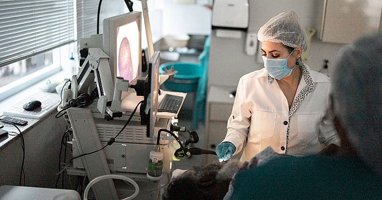
What is Transvaginal Imaging: Overview, Benefits, and Expected Results
The new product is a great addition to our lineup.
Our latest product is an exciting addition to our already impressive lineup! With its innovative features and sleek design, it's sure to be a hit with customers. Don't miss out on this amazing opportunity to upgrade your life!
What is Transvaginal Imaging: Overview, Benefits, and Expected Results
Transvaginal imaging is a diagnostic procedure used to provide detailed images of a female patient’s reproductive organs and pelvic area. It is an important diagnostic tool that provides valuable information for women of childbearing age.
Transvaginal imaging is typically done using transvaginal ultrasonography or transvaginal magnetic resonance imaging (MRI). This article will review the purpose of transvaginal imaging, the benefits of the procedure, and expected results.
What is Transvaginal Imaging
Transvaginal imaging utilizes high-frequency sound waves or magnetic fields to create images of the internal female reproductive organs. During the procedure, a probe is inserted into the patient’s vagina and directed towards the desired area to create the images. Transvaginal ultrasound and MRI are two different procedures that follow the same process.
Transvaginal Ultrasound
A transvaginal ultrasound uses high-frequency sound waves to create images of the internal pelvic structures. The sound waves bounce off the organs and are processed by a computer to create the images. Transvaginal ultrasounds provide a thorough examination of the internal anatomy of the organs and are often used to detect and diagnose a variety of conditions.
Transvaginal Magnetic Resonance Imaging (MRI)
A transvaginal MRI combines magnetic fields, radio waves, and computer technology to generate images of internal pelvic structures. During the procedure, a patient lies on a table in a tube-like device. The patient may be asked to hold a certain position to create the images. The patient may hear loud noises during the procedure.
Benefits of Transvaginal Imaging
Transvaginal imaging is an important diagnostic tool for women of childbearing age. It can provide valuable information about a variety of reproductive issues. The benefits of transvaginal imaging include:
- Identifying the cause of pelvic pain and other reproductive issues
- Detecting reproductive cancer
- Examining the ovaries, uterus, and other organs
- Detecting cysts or tumors
- Evaluating infertility
- Detecting ectopic pregnancies
Transvaginal imaging is a low-risk procedure with minimal discomfort for the patient. The images are generated quickly and can provide valuable information to the patient and doctor.
Expected Results of Transvaginal Imaging
The expected results of transvaginal imaging will vary depending on the reason for the procedure. Generally, the images created will be reviewed by a physician and any abnormalities or issues should be identified. The image results can then be used to help diagnose and treat conditions.
For example, if a patient is having difficulty conceiving, the images from a transvaginal ultrasound can be used to assess the condition of the ovaries, uterus, and other reproductive organs and determine if there are any problems that may be preventing pregnancy.
Conclusion
Transvaginal imaging is an important diagnostic tool for women of childbearing age. It can provide valuable information about a variety of reproductive issues. The procedure is low-risk and the images can be generated quickly, providing valuable insight to the doctor and patient.
The expected results of the procedure will vary depending on the issue being examined. Generally, the images created should provide enough information for the doctor to properly diagnose and treat any reproductive condition.
Transvaginal imaging is a beneficial procedure and can help diagnose and treat a variety of reproductive issues. It is important for women to speak with their doctor if they have any reproductive issues to determine if transvaginal imaging is the right solution.
Definition and Overview
Transvaginal imaging is an ultrasound test used to evaluate the female reproductive system from inside the vagina. Also referred to as endovaginal ultrasound, it is often performed if a woman is suffering from abnormal bleeding and pelvic pain or if polyps, fibroids, and ovarian cysts are suspected. It can also be done in an attempt to identify the cause of female infertility and assess early pregnancy.
Unlike other imaging tests, the procedure uses sound waves and not radiation to create images of the internal organs. Thus, it is safe for the patient or foetus (if the patient is pregnant). It provides better images and more information than traditional pelvic ultrasound because the probe can be positioned closer to the pelvic organs.
Transvaginal ultrasound is performed by sonographers, radiologists, and obstetric sonologists.
Who Should Undergo and Expected Results
A transvaginal ultrasound is used to:
-
Determine the cause of female infertility
-
Determine the cause of heavy and painful periods as well as postmenopausal bleeding
-
Diagnose or confirm ectopic pregnancy
-
Confirm if the patient has uterine fibroids or ovarian cysts
-
Ensure that the intrauterine device (IUD) has been placed properly
-
Investigate the cause of pelvic pain and pain during sexual intercourse
In pregnant women, the procedure is performed to:
-
Monitor the growth of foetus and listen to the baby’s heartbeat
-
Look for signs of abnormalities that can lead to premature delivery or miscarriage
-
Confirm an early pregnancy
-
Detect a possible miscarriage
-
Determine the source of bleeding
-
Diagnose placenta previa or placental abruption. Placenta previa is when the placenta is lying low and covering the cervix. Placental abruption, on the other hand, is when the placenta separates from the uterus before the baby is born.
The test may also be ordered if the results of previous abdominal or pelvic ultrasound suggest an abnormality and to diagnose pelvic infection, ovarian tumours, thickened uterine lining, and birth defects.
The results of the procedure can be immediately interpreted if it is performed by an obstetrician. Otherwise, a technician or a radiologist may send the results to the doctor, who will discuss the findings to the patient on her next appointment.
How is the Procedure Performed?
For the procedure, the patient is asked to remove her clothes from the waist down and put on a gown before lying down on her back with legs spread and knees bent. After covering the ultrasound wand with a condom and lubricating gel, the doctor will insert it into the vagina. Using sound waves, the probe will start to create pictures of the organs, which are immediately transmitted to a nearby monitor.
The procedure is not painful and is thus performed without the use of any anaesthetics. However, there may be some pressure, which many describe as tolerable. It is the same pressure felt during a pap smear when a speculum is inserted into the vagina to collect sample cells from the cervix.
Most patients do not usually need to prepare for the test. However, depending on the reason for the ultrasound, the doctor may need the patient’s bladder to be either partially full or totally empty. The doctor may require the patient to drink about 32 ounces of water prior to the procedure if he needs the intestine to be lifted so clearer pictures of the pelvic organs can be obtained.
If the test is performed to determine whether the endometrium has thickened or if there are polyps, the doctor may also perform saline infusion sonohysterography (SIS) in which a small volume of salt solution is inserted into the uterus so that it can be clearly seen on an ultrasound scan.
Most patients feel normal after the test and are able to resume normal activities right after. However, some experience pelvic discomfort and slight dizziness that commonly last for no more than a few minutes.
Possible Risks and Complications
Transvaginal ultrasound is a safe procedure for both patients and their unborn child (if the patient is pregnant). Aside from slight discomfort during and a couple of minutes after the test, there are no known risks or side effects.
In rare cases where patients are unable to tolerate the procedure, they may opt for a transabdominal ultrasound where the probe is pressed against the abdomen to view the pelvic organs.
References:
-
Transvaginal ultrasound. (n.d.). https://www.cedars-sinai.edu/Patients/Programs-and-Services/Imaging-Center/For-Patients/Exams-by-Procedure/Ultrasound/Transvaginal-Ultrasound.aspx
-
Transvaginal ultrasound: Diagnostics & testing. (2015). http://my.clevelandclinic.org/health/diagnostics/hic-abdominal-renal-ultrasound/hic-transvaginal-ultrasound
/trp_language]
[trp_language language=”ar”][wp_show_posts id=””][/trp_language]
[trp_language language=”fr_FR”][wp_show_posts id=””][/trp_language]


Very informative! #transvaginalimaging