What is a Barium Oesophagram: Overview, Benefits, and Expected Results
Definition & Overview
A barium oesophagram, also referred to as a barium swallow, is a diagnostic procedure used to detect a variety of problems in the digestive system. It requires the patient to swallow a chalk-like mixture called barium. The barium is tracked as it progresses through the upper gastrointestinal tract (GI) using a special x-ray technology. The procedure detects certain abnormalities to help doctors make a definitive diagnosis.
The procedure is usually performed on patients with suspected cases of hiatal hernia, oesophageal diverticula, oesophageal varies, motility disorders, tumours, polyps, or ulcers. In some cases, the procedure may also be performed on patients with gastroesophageal reflux disease (GERD). However, in the recent years, most doctors prefer to perform endoscopy to diagnose and treat this condition.
The upper GI tract is consists of the mouth, throat, oesophagus, stomach, and duodenum. In an endoscopy, the oesophagus, stomach, and duodenum are the main areas of concern. In a barium oesophagram, the entire upper GI tract can be assessed. This means that the procedure can also be useful in diagnosing swallowing difficulties and patients who display the following symptoms:
- Pain in the abdominal area
- Vomit containing blood
- Weight loss for no obvious reasons
- Chest pains
- Sensation of throat blockage
It is important to note that many of the above symptoms can be caused by certain conditions that may prohibit the use of a barium oesophagram. Such conditions include:
- Intestinal obstruction – barium has a tendency to worsen the condition.
- Oesophageal perforation – if barium leaks through a rupture in the oesophagus, it could cause an infection
- Pregnancy – radiation emitted from the x-ray machine can be harmful to pregnant women.
- Poor swallowing reflexes – barium may enter the lungs if the patient has poor swallowing reflexes.
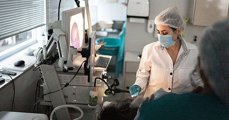
A barium oesophagram makes use of an x-ray imaging device to track the substance as it passes through the GI tract. It is performed by a radiologist who forwards the results to the patient’s attending doctor for a diagnosis.
Who Should Undergo & Expected Results
Barium oesophagram is a highly effective method to search for abnormalities or damages to the upper GI tract. Patients of all ages can undergo the procedure except those with conditions described above.
If the procedure is to be performed on an infant, the child is prevented from eating or drinking for at least 2 hours before the test.
Children between 1 month and 2 years of age may not have anything to eat or drink for at least 4 hours before the test. Children 2 years and older must not eat or drink for at least 8 hours before the test.
A barium oesophagram does not cause any pain or discomfort. However, if the child finds it difficult to drink the barium mixture, a straw may be used. If still not possible, a tube can be inserted into the child’s nose and directed towards the oesophagus. This procedure may cause some discomfort to the child.
The objective of the test is to track the passage of the substance through the upper GI tract. Once this has been accomplished, the test is completed. The entire procedure may take anywhere between 15 and 30 minutes, depending on the condition of the patient.
The images taken during the test are forwarded to the doctor for further examination.
How is the Procedure Performed?
Before a barium oesophagram is performed, the doctor will confirm that the patient is a good candidate for the test. This means that the patient does not have any condition that may worsen due to the mixture.
The doctor then reviews the patient’s detailed medical history and all the symptoms being experienced. If the doctor suspects a problem in the GI tract and determines that a barium oesophagram is the ideal diagnostic procedure, a schedule will be provided to the patient.
The test is performed on a patient with an empty stomach. As such, adults must refrain from eating or drinking anything at least 4 hours before the procedure. If the patient is a child, the conditions stated above will have to be followed.
The radiologist will then prepare the barium mixture and have the patient drink it just before taking images on an x-ray. As mentioned earlier, if the patient is unable to drink the mixture, it can be passed through a flexible tube inserted through the nose and directed towards the oesophagus.
Once the mixture has coated the oesophagus, images will be taken using an x-ray. The radiologist ensures that the images are clear and then submits them to the doctor for further examination.
Possible Risks & Complications
A barium oesophagram is a safe procedure, but it does have associated risks. It is also possible that complications will develop depending on how the procedure was performed or due to the chemical properties of the barium mixture.
The doctor will attempt to determine if the patient is allergic to barium before recommending the procedure. If a flexible tube is used, there is a risk of damage to the upper GI tract. Other risks include constipation or accidental entry of the barium mixture into the trachea.
References:
Children’s Hospital of Pittsburgh;”Eosophagram”; http://www.chp.edu/our-services/radiology/patient-procedures/eosophagram
Cedars-Sinai;”Barium Swallow-Eosophagram”; https://www.cedars-sinai.edu/Patients/Programs-and-Services/Imaging-Center/For-Patients/Exams-by-Procedure/Gastrointestinal-Radiology/Barium-Swallow—Eosophagram.aspx
Jay W. Marks MD; “Upper GI Series (Barium Swallow)”; http://www.medicinenet.com/uppergiseries/article.htm
/trp_language]
[trp_language language=”ar”][wp_show_posts id=””][/trp_language]
[trp_language language=”fr_FR”][wp_show_posts id=””][/trp_language]
## What is a Barium Oesophagram?
**Overview**
A barium oesophagram, also known as a barium swallow, is a medical imaging procedure that evaluates the oesophagus, the hollow muscular tube that carries food from the mouth to the stomach. During the procedure, a thick, white liquid called barium is swallowed, which coats the oesophagus and allows it to be visualized under X-ray or fluoroscopy.
**Benefits**
* **Detects structural abnormalities:** A barium oesophagram can identify narrowing (strictures), blockages, hernias, and other structural issues within the oesophagus.
* **Evaluates function:** By observing the passage of barium through the oesophagus, doctors can assess its motility and ability to move food efficiently.
* **Identifies conditions:** Barium oesophagrams help diagnose gastrointestinal conditions such as:
* Gastroesophageal reflux disease (GERD)
* Barrett’s oesophagus
* Hiatal hernia
* Esophageal cancer
**Expected Results**
* **Normal results:** The oesophagus appears smooth and regular, with no visible abnormalities or delayed emptying.
* **Abnormal results:** Deviations from normal findings may indicate:
* Stricture: Narrowing or blockage of the oesophagus
* Hernia: Displacement of the stomach into the chest cavity
* Motility disorder: Impaired movement of food through the oesophagus
* Tumour or mass: Growth or thickening within the oesophagus
**Additional Information**
* **Preparation:** Fasting is required before the procedure to ensure an empty stomach.
* **Procedure:** The barium swallow involves swallowing a mixture of barium and water. X-ray images are taken to visualize the oesophagus.
* **Radiation:** The amount of radiation exposure is minimal.
* **Follow-up:** Additional tests or treatments may be recommended based on the results of the barium oesophagram.


/* Post title: What is a Barium Oesophagram: Overview, Benefits, and Expected Results */