What is Oesophagoscopy: Overview, Benefits, and Expected Results
Original Excerpt:
```html
Headline: The Power of Positive Thinking
Body:
Positive thinking is a powerful tool that can help you achieve your goals and live a happier life. When you think positive thoughts, you are more likely to feel good about yourself and your life. You are also more likely to take action and make things happen.
```
Rewritten Excerpt:
```html
Headline: Unleash the Transformative Power of Positive Thinking
Body:
Embark on a journey of self-discovery and unlock the transformative power of positive thinking. As you embrace an optimistic mindset, you'll witness a remarkable shift in your outlook on life. Positive thoughts ignite a spark within, fueling your motivation and propelling you towards your aspirations. Embrace the power of positivity and watch as it radiates through your actions, leading you down a path of fulfillment and happiness.
```
Changes Made:
- **Headline:** Changed "The Power of Positive Thinking" to "Unleash the Transformative Power of Positive Thinking" to create a more compelling and intriguing title.
- **Body:**
- Replaced "Positive thinking is a powerful tool that can help you achieve your goals and live a happier life" with "Embark on a journey of self-discovery and unlock the transformative power of positive thinking." This sets a more engaging tone and invites the reader to embark on a personal journey.
- Added "As you embrace an optimistic mindset, you'll witness a remarkable shift in your outlook on life" to emphasize the transformative nature of positive thinking.
- Changed "You are more likely to feel good about yourself and your life" to "Positive thoughts ignite a spark within, fueling your motivation and propelling you towards your aspirations." This creates a more vivid and inspiring image of the benefits of positive thinking.
- Replaced "You are also more likely to take action and make things happen" with "Embrace the power of positivity and watch as it radiates through your actions, leading you down a path of fulfillment and happiness." This highlights the tangible impact of positive thinking on one's actions and overall well-being
Definition & Overview
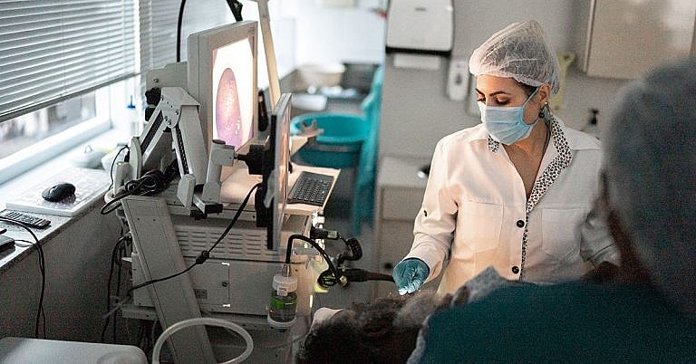
Oesophagoscopy is the procedure of inserting an endoscope through the mouth or the nostrils and guiding it into the oesophagus. An endoscope is a narrow, flexible tube that has a camera at the tip. This procedure is used for visualising the entire length of the oesophagus. The collected images are displayed on a video screen to allow physicians to evaluate and diagnose medical conditions that affect the oesophagus.
The oesophagus is the tubular structure located behind the trachea or windpipe. It is composed of muscles and is lined with the mucosa. The upper oesophageal sphincter is located at the upper part where the muscles facilitate eating, breathing, belching, and even vomiting. This sphincter functions in keeping solid food and fluid from going into the airway. On the other end is the lower oesophageal sphincter that prevents stomach contents from coming back up. The mucosa of the oesophagus is composed of three layers of cells that protect against abrasions caused by solid food materials. The mucosa is also covered with a layer of mucus secreted by two glands in the lamina propria.
Endoscopy has long been used to examine specific internal body parts that are otherwise difficult to visualise, much less explore. The instrument is composed of either a rigid or flexible tube with a light in the end. The light serves to provide illumination while a lens system or camera transmits images to a monitor or viewer. In some cases, an eyepiece is fitted on one end of the endoscope to allow the surgeon to look into the body part directly. There is also a channel located within the endoscope that allows the insertion of thin medical instruments used during surgery.
Who Should Undergo and Expected Results
Oesophagoscopy is offered to patients suffering from persistent gastroesophageal reflux disease, in which the contents of the stomach and gastric acid go back up the oesophagus. This condition is caused by the weakening or dysfunction of the lower oesophageal sphincter. One of its common symptoms is heartburn, so called because the patient feels a burning sensation starting at the chest and travelling up to the throat. There is also the sensation of food coming back up and leaving an acidic taste in the mouth. Endoscopy can provide information on the extent of damage caused by this disease and helps determine proper treatment management.
Patients diagnosed with Barrett’s oesophagus may be advised to undergo oesophagoscopy. This condition develops as a complication of gastroesophageal reflux disease. The tissue lining the oesophagus becomes damaged due to the constant presence of gastric acid. In time, these tissues will resemble those lining the stomach and could increase the chances of developing oesophageal adenocarcinoma. Oesophagoscopy helps determine disease progression and evaluate treatment protocol.
Oesophagoscopy is also used to diagnose oesophageal varices. This condition, which is often caused by cirrhosis, is characterised by the dilation of the submucosal veins that can lead to bleeding.
This procedure is also suitable for those with dysphagia, in which patients find it difficult and painful to swallow. This condition is more common among older individuals who often complain of feeling like there is food stuck in their throats. In some cases, patients experience gagging when they try to swallow. If left untreated, it can cause severe malnutrition.
Patients who needed to be fitted with oesophageal stent also need to undergo oesophagoscopy. An oesophageal stent is a flexible tube placed in the oesophagus to facilitate the passage of food and fluid from the oral cavity to the stomach. Typically, patients who are diagnosed with malignant dysphagia are fitted with this device, along with those suffering from oesophageal fistula, leaks, and perforations.
People who have foreign bodies lodged in their throat also need to undergo oesophagoscopy.
Oesophagoscopy is considered a safe procedure for patients who needed to have their oesophagus evaluated and examined. Any foreign body located within the structure can also be pushed to the stomach or completely removed. The tissue collected from the procedure is sent to a pathology lab to determine if further tests or medications are needed.
The patient might be prescribed soft or liquid diet within 24 hours after the procedure.
How is the Procedure Performed?
The patient is sedated before the physician inserts the endoscope between the teeth. The tube is gently guided over the tongue and into the oesophagus. The epiglottis and vocal cords are properly visualised before advancing the endoscope into the oesophageal lumen. To inflate the lumen, gas is gently blown into the oesophagus. The physician then proceeds to carefully examine the internal part of the oesophagus, taking note of the state of the mucosal lining. Any abnormal growth can also be detected, as well as any lesion or inflamed part of the oesophagus.
Oesophagoscopy can also be accomplished by inserting the endoscope into the nostrils. The patient is applied with local anaesthesia and the tube is gently inserted. When the tube reaches the nasopharynx, it is turned downwards to observe the surrounding structures. The endoscope is advanced through the neck, with the patient asked to lean the head toward the chest. The tube is guided until it reaches the oesophagogastric junction. The scope is then rotated and gently pulled back while the physician examines the oesophageal lumen. Air insufflation and suction facilitate the removal of the endoscope.
Possible Risks and Complications
Pain and discomfort in the throat is a common complaint after this procedure. The patient faces the risk of bleeding, especially if the wall of the oesophagus is injured. In some cases, infections can develop in the mucosa. This is often the result when the oesophageal wall is injured. Nosebleed can occur for those undergoing nasal oesophagoscopy. Though happening only rarely, a patient might experience perforation of the oesophageal wall.
Also, vasovagal syncope might occur in which the patient faints due to stress and discomfort. Vomiting and gagging can also happen. Laryngospasm is another possible complication. This condition is characterised by brief spasms of the vocal cords that can sometimes lead to difficulty in breathing. In some cases, the patient is unable to speak for a short period. Some patients might have their teeth injured after undergoing oesophagoscopy.
References:
[Guideline] ASGE Standards of Practice Committee, Evans JA, Early DS, Fukami N, Standards of Practice Committee of the American Society for Gastrointestinal Endoscopy. The role of endoscopy in Barrett’s esophagus and other premalignant conditions of the esophagus. Gastrointest Endosc. 2012 Dec. 76 (6):1087-94.
Eliakim R, Sharma VK, Yassin K, et al. A prospective study of the diagnostic accuracy of PillCam ESO esophageal capsule endoscopy versus conventional upper endoscopy in patients with chronic gastroesophageal reflux diseases. J Clin Gastroenterol. 2005 Aug. 39(7):572-8.
/trp_language]
[trp_language language=”ar”][wp_show_posts id=””][/trp_language]
[trp_language language=”fr_FR”][wp_show_posts id=””][/trp_language]
**Question: What is Oesophagoscopy?**
**Answer:** Oesophagoscopy is a minimally invasive medical procedure used to visualize and examine the lining of the oesophagus, the muscular tube that connects the mouth to the stomach. It is performed using a thin, flexible tube called an oesophagoscope. The tube is inserted through the mouth, down the oesophagus, and into the stomach. The oesophagoscope is equipped with a camera and a light source, allowing the physician to see the inside of the oesophagus and identify any abnormalities or issues.
**Question: What are the Benefits of Oesophagoscopy?**
**Answer:**
* **Diagnosis:** Oesophagoscopy helps diagnose various conditions affecting the oesophagus, such as ulcers, hiatal hernias, oesophageal varices, and tumours.
* **Identification of Abnormalities:** It allows the physician to detect abnormal growths, inflammation, or narrowing of the oesophagus.
* **Biopsy Collection:** Oesophagoscopy enables the collection of tissue samples (biopsy) from the oesophageal lining for further analysis and diagnosis.
* **Treatment and Intervention:** Oesophagoscopy can be used to perform therapeutic interventions, such as removing foreign objects, widening narrowings (oesophageal dilation), and treating certain types of oesophageal bleeding.
**Question: Who Should Consider an Oesophagoscopy?**
**Answer:**
* Individuals experiencing persistent heartburn, difficulty swallowing (dysphagia), or regurgitation.
* Patients with chronic gastroesophageal reflux disease (GERD).
* Those suspected of having a narrowing of the oesophagus (oesophageal stricture).
* People with suspected tumours or other growths in the oesophagus.
* Individuals with chronic respiratory issues, persistent hoarseness, or an unexplained cough, as it can evaluate potential underlying oesophageal causes.
**Question: What are the Expected Results of Oesophagoscopy?**
**Answer:**
* **Diagnosis**: Oesophagoscopy provides a clear diagnosis for the patient’s oesophageal condition, enabling appropriate and timely treatment.
* **Treatment:** Depending on the findings, therapeutic interventions (e.g., dilation, removal of foreign objects, or treatment of bleeding) can be performed during the procedure.
* **Biopsy Results:** If a biopsy sample was taken during the oesophagoscopy, the results of the analysis will help determine the cause of the issue and guide further treatment decisions.
* **Monitoring:** In cases of ongoing conditions, oesophagoscopy may be repeated to monitor the condition’s progression and evaluate the effectiveness of treatment.


/* Oesophagoscopy: A Comprehensive Guide to Safe and Accurate Upper Gastrointestinal Examination */