
Arteriovenous Malformation (AVM) Surgery: Comprehensive Overview
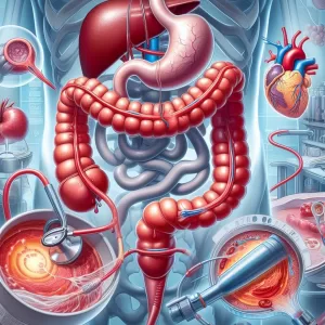
Three kinds of surgery are capable of treating an arteriovenous malformation, a tangle of blood vessels that impedes blood flow.
 Surgery: Comprehensive Overview 1")
An arteriovenous malformation (AVM) is a rare, noncancerous entanglement of blood vessels that prevents blood from flowing between your arteries and veins.
Instead of blood flowing from your arteries through capillaries to your veins, an AVM causes blood to flow directly from the arteries into the veins. Your capillaries are unable to distribute oxygen-rich blood to cells, which may damage them.
The excessive blood circulating in an AVM may lead to bleeding, especially for young adults. This could cause neurological problems, like seizures or strokes, or may be fatal.
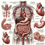
An AVM can develop anywhere in your body. AVMs in the brain or spinal cord are especially concerning due to the bleeding risk. Because only about 12%
AVMs have symptoms; many are discovered during brain imaging tests for another condition.
For most AVMs, surgery is needed to remove them or reduce their size.
Read on to learn about the types of surgery for an AVM and what to expect for each type.
AVM surgery purpose
The purpose of AVM surgery is to prevent the possibility of bleeding by removing or reducing the tangled blood vessels.
This allows blood to flow normally through your capillaries.
AVM surgery types
The following three types of surgery can treat AVMs:. A doctor determines the best option based on factors including:
Radiochirurgie stéréotaxique
Stereotactic radiosurgery uses focused radiation beams to thicken and close the AVM’s blood vessels without destroying healthy tissue.
It’s the least invasive surgery to treat an AVM. It doesn’t require an incision. This painless procedure uses sophisticated equipment like a Gamma Knife or CyberKnife.
Stereotactic radiosurgery may be used for AVMs that are difficult to reach or alongside surgical removal or endovascular embolization.
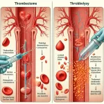
Endovascular embolization
This less invasive type of surgery can only be performed for AVMs that can be reached with a catheter, which is a long, thin tube.
A surgeon inserts the catheter in an artery, guides it to the AVM, and injects materials that stop blood from flowing to the AVM.
Surgical removal (resection)
Surgical removal, or resection, is the only surgery type that completely removes the AVM. It’s the most invasive and riskiest type. Doctors generally only recommend it for an AVM that has ruptured or has a high chance of rupturing.
To lower the risk of complications, a doctor may first use endovascular embolization or radiosurgery to shrink the AVM, making it safer to remove.
AVM surgery: side effects and risks
All types of AVM surgery may have the following side effects and risks:
Bleeding in the brain may occur during the 24 hours after AVM surgery. According to 2021 research
Out of 214 AVM surgeries, two cases of bleeding began at least 24 hours after surgery.
Additionally, each type of surgery may have additional effects:
- Radiosurgery:
- bruising, tenderness, or temporary hair loss where a metal plate was attached to your scalp
- swelling in the treated area
- tenderness
- Endovascular embolization:
- stroke-like symptoms, such as weakness in one arm or leg
- Surgical removal:
- soreness, numbness, or itchiness around the incision
- swelling or bruising around your eyes
AVM surgery survival rate
Adverse outcomes for AVM surgery range from 10% for smaller AVMs to more than 30% for larger ones, according to a 2023 review.
In addition to the AVM size, factors affecting the survival rate include its location and your age.
AVM surgery procedures
The three types of AVM surgery range from radiosurgery, which is noninvasive, to resection, which is the most invasive type.
Stereotactic radiosurgery procedure
Stereotactic radiosurgery may take several hours. You may need to repeat this treatment two or three times over the course of a few months.
The steps are generally as follows:
Endovascular embolization procedure
This procedure may be performed while you’re asleep under general anesthesia, or you may remain awake and receive medication to relax you. It may take 3–8 hours.
The steps are generally as follows:
A similar procedure called sclerotherapy may be used to treat AVMs. It also uses a catheter, but unlike endovascular embolization, which blocks blood flow with injected materials, sclerotherapy uses an injection sclerosant. This medication destroys the AVM’s blood vessels.
Resection procedure
For a resection procedure, general anesthesia is used, so you’re asleep during the procedure.
The steps are generally as follows:
Preparing for AVM surgery
Speak with your doctor about any medications you’re taking and what medications you can take on the day of the surgery.
You will usually be asked not to eat or drink anything for 8 hours before surgical removal or endovascular embolization.
Before surgical removal of an AVM, the hair around the incision site is shaved and your scalp cleaned. For radiotherapy, you may need to use a special shampoo before the procedure. Do not use hairspray or gel.
For endovascular embolization and radiotherapy, wear comfortable, loose clothing.
For radiotherapy, do not wear:
AVM surgery recovery
The recovery depends on the type of AVM surgery.
After AVM surgical removal that has no complications, you’ll probably stay in the hospital for 4–6 days and spend at least the first day in the intensive care unit (ICU).
If bleeding occurs during surgery, you may need to stay longer in the hospital.
Following an endovascular embolization, you may need to stay in the ICU for 1 day or longer if bleeding occurs.
You’re usually able to go home after radiosurgery, but do plan on having someone drive you to and from your appointment. The medication used during radiosurgery can make you sleepy.
AVM surgery recovery time
You can typically resume your daily activities 4–6 weeks after the surgical removal of an AVM. It may take up to 6 months to fully recover.
After endovascular embolization or radiosurgery, you can generally resume your normal activities the day after your procedure.
How effective is AVM surgery?
In a small 2021 study
With 44 people whose AVMs were surgically removed, researchers found that 88% of them had a good outcome 3 months later.
Five people experienced a moderate disability associated with bleeding. One person experienced a severe disability.
According to a 2022 review
Of the 36 studies that included 2,108 people who had endovascular embolization, 80% experienced obliteration (destruction) of their AVMs on average.
It may take about 3 years to completely destroy an AVM with radiosurgery. About 70–80% of AVMs are destroyed during this time frame.
AVM surgery cost
Although there’s not much data available on the cost of treatment for AVMs, a 2019 study
with 140 people with AVMs who were treated from 2012–2015 found the following average costs:
Several factors determine the cost, including the size of the AVM and whether it has ruptured.
Health insurance and federal government programs may cover some of the cost of AVM surgery. Contact your insurance provider for information specific to your situation.
Frequently asked questions about AVM surgery
The following are answers to some frequently asked questions regarding AVM surgery.
How serious is AVM surgery?
Surgical removal of an AVM is considered high risk
due to its location in the brain or spine and the possibility of serious complications.
Endovascular embolization and radiotherapy are less invasive and have fewer risks.
What are the chances of surviving an AVM?
It’s possible to live with an AVM. However, bleeding causes temporary or permanent brain damage in 20–30% of cases and death in 10–15% of cases, according to the Brain Aneurysm Foundation.
What is the AVM life expectancy?
The life expectancy of someone with an AVM depends on several factors, such as whether it ruptures, its size and location, the person’s age, and the effectiveness of treatment.
Emporter
The only way to completely remove an AVM is through resection, but it’s a high-risk surgery. Doctors usually recommend it only for AVMs that have ruptured or have the potential to rupture.
Less invasive surgeries are endovascular embolization and radiosurgery. They both reduce the size of the AVM and may obliterate it over time.
A doctor can recommend the surgery that’s best for your particular case.
Un commentaire
Laisser un commentaire
Articles Similaires
Articles populaires


AVM surgery: What you need to know